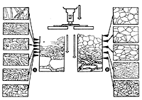
Mantle preparations should be done after the descriptions published by Agerer (1991): Methods in Microbiology 23: 25-73. A Normarski interference contrast microscope (DIC) is most convenient to examine the mantles, since it allows to focus different mantle layers consecutively, without interference from hyphal layers laying beneath or above the focused level (A). One part of the mantle pieces should be oriented with the outer side up, another part with the inner side up. Only very dark mantles, or mantles covered by many strongly light reflecting crystals or soil particles need to be sectioned; these sections can be made as thick tangential sections (it is enough to halve the mantle) by the aid of a cryotome. It is necessary to always apply the highest magnification (1000×); only for discerning patterns of hyphal arrangement, lower magnifications should be used (400×). The application of a well adjusted microscope is self-evident.
With some experience it is easy to distinguish between inner mantle and outer mantle surface. Hints for an outer mantle surface are:
- a convex side or/and
pseudoparenchymatous structure or/and
soil particles or/and
crystals or/and
emanating elements or/and
dark cell walls.
Hints for an inner mantle surface:
- concave side or/and
plectenchymatous structure or/and
root cell wall remnants
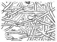
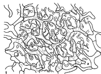
The structure of the outer mantle layer, frequently called mantle surface, is the most informative portion of the mantle. In some cases, it is difficult to define exactly the mantle surface, particularly, when a hyphal mantle is loosely woven (B) or when a proper mantle is covered by hyphae which can either be regarded as parts of the mantle or as parts of the emanating elements (C).
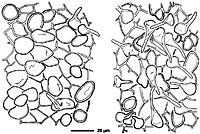
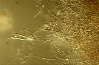
Generally, in cases where a hyphal net (D) or similar structures are formed which are intimately associated with the proper mantle (E), these structures are regarded as being a part of the mantle surface. In loosely plectenchymatous mantles, the mantle surface is considered as it appears in a microscope slide after a piece of mantle is covered by a cover slip; most of the time, the proximal parts of emanating hyphae are included, distal parts are frequently spread out in the neighbourhood of the mantle piece (F).
 A |
 B |
 C |
 D |
 E |
 F |