|
|
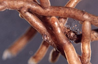
– Enlarged view – |
| • references | |
| Gronbach E (1988) Charakterisierung und Identifizierung von Ektomykorrhizen in einem Fichtenbestand mit Untersuchungen zur Merkmalsvariabilität in sauer beregneten Flächen. Bibl Mycol 125: 1-217. Gronbach E (1989) Picierhiza conspicua. In Agerer R (ed) Colour Atlas of Ectomycorrhizae, plate 29, Einhorn-Verlag, Schwäbisch Gmünd. |
|
| • length | |
| 3 mm | Lower value of unspecified range (could be µ-s.d., but not known) |
| 22 mm | Upper value of unspecified range (could be µ+s.d., but not known) |
| • ramification presence-type | |
| monopodial-pyramidal | |
| • tips {per 10 mm} number | |
| 8 | Lower value of unspecified range (could be µ-s.d., but not known) |
| 10 | Upper value of unspecified range (could be µ+s.d., but not known) |
| • ramification orders | |
| 0 | Lower value of unspecified range (could be µ-s.d., but not known) |
| 2 | Upper value of unspecified range (could be µ+s.d., but not known) |
| • main axis diameter | |
| 0.4 mm | Lower value of unspecified range (could be µ-s.d., but not known) |
| 0.5 mm | Upper value of unspecified range (could be µ+s.d., but not known) |
| • rhizomorphs as stout, short, conical structures presence-abundance | |
| absent | |
| • rhizomorphs as short mycorrhiza-like outgrowths with blunt tips presence | |
| absent | |
| • rhizomorphs presence | |
| present | |
| • rhizomorphs frequency | |
| infrequent | |
| • shape | |
| straight | |
| or | bent |
| or | beaded |
| • shape {of distal end} | |
| not inflated, cylindric | |
| • length | |
| 0 mm | Lower value of unspecified range (could be µ-s.d., but not known) |
| 4 mm | Upper value of unspecified range (could be µ+s.d., but not known) |
| • diameter | |
| 0.3 mm | Lower value of unspecified range (could be µ-s.d., but not known) |
| 0.4 mm | Upper value of unspecified range (could be µ+s.d., but not known) |
| • colour | |
| ochre, yellowish brown | |
| • very tip colour | |
| ochre, yellowish brown | |
| or | whitish |
| • older parts colour | |
| dark brown | |
| or | brown |
| • mantle cortical cells visibility | |
| not visible | |
| or | visible |
| • mantle {distinct} surface visibility | |
| present | |
| • mantle laticifers visibility | |
| absent | |
| • mantle dots presence-colour | |
| absent | |
| • mantle carbonizing presence | |
| absent | |
| • mantle surface {in general} habit | |
| shiny | |
| or | smooth |
| • emanating hyphae presence | |
| present | |
| • emanating hyphae abundance | |
| infrequent | |
| • presence | |
| absent | |
| • emanating elements presence-type | |
| rhizomorphs | |
| or | cystidia |
| • emanating elements cystidia location | |
| on outer mantle layer | |
| • presence | |
| absent | |
| • organisation | |
| plectenchymatous | |
| • mantle type | |
| hyphae rather irregularly arranged and no special pattern discernible (type B) | |
| • hyphal system kind | |
| with rather short, obtuse, even finger-like branches | |
| • cell diameter | |
| 2 µm | Lower value of unspecified range (could be µ-s.d., but not known) |
| 3 µm | Upper value of unspecified range (could be µ+s.d., but not known) |
| 5 µm | Maximum value |
| • cell length | |
| 10 µm | Lower value of unspecified range (could be µ-s.d., but not known) |
| 40 µm | Upper value of unspecified range (could be µ+s.d., but not known) |
| • cell wall thickness | |
| 0.2 µm | Lower value of unspecified range (could be µ-s.d., but not known) |
| 0.5 µm | Upper value of unspecified range (could be µ+s.d., but not known) |
| • drops of exuded pigment presence | |
| absent | |
| • organisation | |
| plectenchymatous | |
| • hyphae arrangement | |
| plectenchymatous, ring-like | |
| • cell diameter | |
| 2 µm | Lower value of unspecified range (could be µ-s.d., but not known) |
| 3 µm | Upper value of unspecified range (could be µ+s.d., but not known) |
| 5 µm | Maximum value |
| • cell length | |
| 10 µm | Lower value of unspecified range (could be µ-s.d., but not known) |
| 40 µm | Upper value of unspecified range (could be µ+s.d., but not known) |
| • cell wall thickness | |
| 0.2 µm | Lower value of unspecified range (could be µ-s.d., but not known) |
| 0.5 µm | Upper value of unspecified range (could be µ+s.d., but not known) |
| • cell wall surface habit | |
| smooth | |
| • organisation | |
| plectenchymatous | |
| • hyphae arrangement | |
| with some considerably inflated cells | |
| • septa clamps presence | |
| present | |
| • cell diameter | |
| 2 µm | Lower value of unspecified range (could be µ-s.d., but not known) |
| 3 µm | Upper value of unspecified range (could be µ+s.d., but not known) |
| 5 µm | Maximum value |
| • cell length | |
| 10 µm | Lower value of unspecified range (could be µ-s.d., but not known) |
| 40 µm | Upper value of unspecified range (could be µ+s.d., but not known) |
| • anatomy mantle outer mantle layer {of ectomycorrhizal tip} organisation | |
| plectenchymatous | |
| or | pseudoparenchymatous |
| • anatomy mantle outer mantle layer {of ectomycorrhizal tip} hyphae diameter | |
| 2 µm | Lower value of unspecified range (could be µ-s.d., but not known) |
| 6 µm | Upper value of unspecified range (could be µ+s.d., but not known) |
| • anatomy mantle outer mantle layer {of ectomycorrhizal tip} cell density | |
| 10 | Lower value of unspecified range (could be µ-s.d., but not known) |
| 14 | Upper value of unspecified range (could be µ+s.d., but not known) |
| • mantle thickness {apart from tip} | |
| 10 µm | Lower value of unspecified range (could be µ-s.d., but not known) |
| 45 µm | Upper value of unspecified range (could be µ+s.d., but not known) |
| 80 µm | Maximum value |
| • mantle different layers presence | |
| not discernable | |
| • outer mantle layer organisation | |
| plectenchymatous | |
| • middle mantle layer organisation | |
| plectenchymatous | |
| • inner mantle layer organisation | |
| plectenchymatous | |
| • unlayered mantle hyphae tangentially length | |
| 2 µm | Lower value of unspecified range (could be µ-s.d., but not known) |
| 7 µm | Upper value of unspecified range (could be µ+s.d., but not known) |
| 20 µm | Maximum value |
| • unlayered mantle hyphae radially diameter | |
| 2 µm | Lower value of unspecified range (could be µ-s.d., but not known) |
| 5 µm | Upper value of unspecified range (could be µ+s.d., but not known) |
| 7 µm | Maximum value |
| • presence | |
| present | |
| • shape | |
| tangentially-oval, -elliptic or -cylindrical, and oriented in parallel to root axis | |
| • tangentially length | |
| 75 µm | Lower value of unspecified range (could be µ-s.d., but not known) |
| 120 µm | Upper value of unspecified range (could be µ+s.d., but not known) |
| 180 µm | Maximum value |
| • radially diameter | |
| 7 µm | Minimum value |
| 10 µm | Lower value of unspecified range (could be µ-s.d., but not known) |
| 35 µm | Upper value of unspecified range (could be µ+s.d., but not known) |
| 45 µm | Maximum value |
| • mean tangenial length TCt | |
| 98.4 µm | Mean (= average) |
| • mean shape-ratio TCq | |
| 3.8 | Mean (= average) |
| • anatomy mantle longitudinal section cortical (epidermal) cells shape | |
| tangentially-oval to -elliptic or -cylindrical, and oriented in parallel to root axis | |
| • anatomy mantle longitudinal section cortical (epidermal) cells tangentially length | |
| 90 µm | Lower value of unspecified range (could be µ-s.d., but not known) |
| 130 µm | Upper value of unspecified range (could be µ+s.d., but not known) |
| 140 µm | Maximum value |
| • anatomy mantle longitudinal section cortical (epidermal) cells radially diameter | |
| 12 µm | Minimum value |
| 15 µm | Lower value of unspecified range (could be µ-s.d., but not known) |
| 35 µm | Upper value of unspecified range (could be µ+s.d., but not known) |
| 45 µm | Maximum value |
| • anatomy mantle longitudinal section cortical (epidermal) cells mean tangential length CCt (ECt) | |
| 108.2 µm | Mean (= average) |
| • anatomy mantle longitudinal section cortical (epidermal) cells mean shape-ratio CCq (ECq) | |
| 3.9 | Mean (= average) |
| • presence | |
| present | |
| • kind | |
| protruding towards endodermis | |
| • structure {in plan view} | |
| of palmetti type | |
| • mantle different layers presence | |
| not discernible | |
| • outer mantle layer organisation | |
| plectenchymatous | |
| • middle mantle layer organisation | |
| plectenchymatous | |
| • inner mantle layer organisation | |
| plectenchymatous | |
| • unlayered mantle hyphae tangentially length | |
| 2 µm | Lower value of unspecified range (could be µ-s.d., but not known) |
| 7 µm | Upper value of unspecified range (could be µ+s.d., but not known) |
| 20 µm | Maximum value |
| • unlayered mantle hyphae radially diameter | |
| 2 µm | Lower value of unspecified range (could be µ-s.d., but not known) |
| 5 µm | Upper value of unspecified range (could be µ+s.d., but not known) |
| 7 µm | Maximum value |
| • presence | |
| present | |
| • rows number | |
| 1 | Lower value of unspecified range (could be µ-s.d., but not known) |
| 2 | Upper value of unspecified range (could be µ+s.d., but not known) |
| • shape | |
| tangentially-oval to tangentially-elliptic | |
| • tangentially length | |
| 25 µm | Lower value of unspecified range (could be µ-s.d., but not known) |
| 60 µm | Upper value of unspecified range (could be µ+s.d., but not known) |
| • radially diameter | |
| 7 µm | Minimum value |
| 10 µm | Lower value of unspecified range (could be µ-s.d., but not known) |
| 35 µm | Upper value of unspecified range (could be µ+s.d., but not known) |
| 45 µm | Maximum value |
| • mean tangential length TCt | |
| 40.6 µm | Mean (= average) |
| • mean shape-ratio TCq | |
| 2.9 | Mean (= average) |
| • anatomy mantle cross-section cortical (epidermal) cells shape | |
| tangentially-oval to tangentially-elliptic | |
| • anatomy mantle cross-section cortical (epidermal) cells tangentially length | |
| 15 µm | Minimum value |
| 20 µm | Lower value of unspecified range (could be µ-s.d., but not known) |
| 45 µm | Upper value of unspecified range (could be µ+s.d., but not known) |
| • anatomy mantle cross-section cortical (epidermal) cells radially diameter | |
| 12 µm | Minimum value |
| 15 µm | Lower value of unspecified range (could be µ-s.d., but not known) |
| 35 µm | Upper value of unspecified range (could be µ+s.d., but not known) |
| 45 µm | Maximum value |
| • anatomy mantle cross-section cortical (epidermal) cells mean tangential length CCt | |
| 32.1 µm | Mean (= average) |
| • anatomy mantle cross-section cortical (epidermal) cells mean shape-ratio CCq | |
| 1.8 | Mean (= average) |
| • presence | |
| present | |
| • kind | |
| protruding towards endodermis | |
| • anatomy mantle cross-section hyphal cells around tannin cells shape | |
| roundish | |
| • anatomy mantle cross-section hyphal cells around tannin cells thickness | |
| 2 µm | Lower value of unspecified range (could be µ-s.d., but not known) |
| 10 µm | Upper value of unspecified range (could be µ+s.d., but not known) |
| • anatomy mantle cross-section hyphal rows around tannin cells number | |
| one | |
| or | two |
| • anatomy mantle cross-section hyphal cells around cortical (epidermal) cells shape | |
| roundish | |
| • anatomy mantle cross-section hyphal cells around cortical (epidermal) cells thickness | |
| 2 µm | Lower value of unspecified range (could be µ-s.d., but not known) |
| 4 µm | Upper value of unspecified range (could be µ+s.d., but not known) |
| • anatomy mantle cross-section hyphal rows around cortical (epidermal) cells number | |
| one | |
| • structure {in plan view} | |
| of palmetti type | |
| • intrahyphal hyphae presence | |
| present | |
| • septal pores configuration | |
| globular thickenings | |
| • anastomoses type | |
| open, with a short bridge or bridge almost lacking | |
| • anastomoses cell wall thickness {relative to remaining cell walls} | |
| as thick as | |
| • anastomoses location | |
| not specified | |
| • type | |
| awl-shaped, bristle-like (type A) | |
| • ramification presence-position | |
| absent | |
| • cell wall colour | |
| absent | |
| • cell wall thickness {relative to mantle cells} | |
| thicker | |
| • cell wall evenness | |
| even in thickness | |
| • surface habit | |
| smooth | |
| • shape | |
| not striking | |
| • cell pigment location-colour | |
| absent | |
| • drops of exuded pigment presence | |
| absent | |
| • clamps presence | |
| present | |
| • clamps outline {in lateral view} | |
| less than a semicircle | |
| or | not constricted at contact point to subtending hyphal cell |
| • clamps width {relative to hypha in lateral view} | |
| thinner than | |
| • clamps hole presence | |
| absent | |
| • clamps blister-like structure {at basis} presence | |
| absent | |
| • presence | |
| present | |
| • abundance | |
| infrequent | |
| • distribution | |
| not specified | |
| • anatomy emanating elements emanating hyphae cell shape | |
| with ellbow-like protrusions | |
| • anatomy emanating elements emanating hyphae cell diameter | |
| 2.5 µm | Lower value of unspecified range (could be µ-s.d., but not known) |
| 5 µm | Upper value of unspecified range (could be µ+s.d., but not known) |
| 7 µm | Maximum value |
| • anatomy emanating elements emanating hyphae cell length | |
| 10 µm | Lower value of unspecified range (could be µ-s.d., but not known) |
| 120 µm | Upper value of unspecified range (could be µ+s.d., but not known) |
| • anatomy emanating elements emanating hyphae cell wall surface habit | |
| smooth | |
| or | without lens-shaped appositions |
| or | without spindle-shaped appositions |
| • anatomy emanating elements emanating hyphae cell wall thickness | |
| 0.2 µm | Lower value of unspecified range (could be µ-s.d., but not known) |
| 1.3 µm | Upper value of unspecified range (could be µ+s.d., but not known) |
| 2 µm | Maximum value |
| • anatomy emanating elements emanating hyphae cell wall {apart from tip} evenness | |
| uneven in thickness | |
| • type | |
| undifferentiated; hyphae rather loosely woven and of uniform diameter (type A) |
|
| • internal nodia presence | |
| absent | |
| • gelatinized hyphae presence | |
| present | |
| • cup-like structures on surface presence | |
| absent | |
| • a "ball" of intertwined, ramified, thin hyphae presence | |
| absent | |
| • ampullate, trumpet-like inflated presence | |
| absent | |
| • anatomy emanating elements rhizomorphs hyphae ampullate, trumpet-like inflated presence | |
| absent | |
| • anatomy emanating elements rhizomorphs hyphae ampullate, trumpet-like inflated presence | |
| absent | |
| • anatomy emanating elements rhizomorphs hyphae ampullate, trumpet-like inflated presence | |
| absent | |
| • anatomy emanating elements rhizomorphs hyphae ampullate, trumpet-like inflated presence | |
| absent | |
| • presence | |
| absent | |
| • {of ectomycorrhiza former} presence | |
| present | |
| • {of ectomycorrhiza former} abundance | |
| occasionally present | |
| • shape | |
| as normal hyphae | |
| or | globular |
| • number {per cell} | |
| 1 | Minimum value |
| 2 | Mean (= average) |
| 3 | Maximum value |
| • shape | |
| round | |
| • diameter | |
| 1 µm | Lower value of unspecified range (could be µ-s.d., but not known) |
| 1.5 µm | Upper value of unspecified range (could be µ+s.d., but not known) |
| 2 µm | Maximum value |
| • length | |
| 1 µm | Lower value of unspecified range (could be µ-s.d., but not known) |
| 1.5 µm | Upper value of unspecified range (could be µ+s.d., but not known) |
| 2 µm | Maximum value |
| • geographic occurrence continent | |
| Europe | |
| • plant family | |
| Pinaceae | |
| • plant genus | |
| Picea | |
| • plant habitat kind | |
| forests, woods | |
| • public notes | |
| Mycorrhizal systems irregularly monopodial- pyramidal; mycorrhizal ends almost straight, in part beaded; very tips lighter coloured to hyaline; plan view of outer mantle layer of very tips showing cells which appear epidermoid or irregularly oval to roundish; cystidia with a foot-cell; cell walls of emanating hyphae sometimes appearing multi- layered; autofluorescence of mantle with UV-filter whitish to blue-green, with blue-filter light yellow to ochre; mantle in sulfo-vanillin with slightly rosy walls, in FeSO4 walls slightly yellow-green, in cotton-blue walls bluish, in acid fuchsin walls slightly rosy. | |