|
|
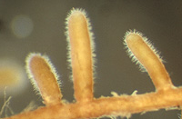
– Enlarged view – |
| • references | |
| Blaschke H (1987) Vorkommen und Charakterisierung der Ektomykorrhizaassoziation Tuber puberulum mit Picea abies. Z Mykol 53: 283-288. Blaschke H (1988) Tuber puberulum. In Agerer R (ed) Colour Atlas of Ectomycorrhizae, plate 22, Einhorn-Verlag, Schwäbisch Gmünd. |
|
| • length | |
| 1 mm | Lower value of unspecified range (could be µ-s.d., but not known) |
| 5 mm | Upper value of unspecified range (could be µ+s.d., but not known) |
| • ramification presence-type | |
| monopodial-pyramidal | |
| • ramification orders | |
| 0 | Lower value of unspecified range (could be µ-s.d., but not known) |
| 1 | Upper value of unspecified range (could be µ+s.d., but not known) |
| • abundance | |
| solitary or in small numbers | |
| • rhizomorphs as stout, short, conical structures presence-abundance | |
| absent | |
| • rhizomorphs as short mycorrhiza-like outgrowths with blunt tips presence | |
| absent | |
| • rhizomorphs presence | |
| absent | |
| or | present |
| • rhizomorphs frequency | |
| infrequent | |
| • exploration type | |
| short distance | |
| • shape | |
| straight | |
| • shape {of distal end} | |
| not inflated, cylindric | |
| • diameter | |
| 0.4 mm | Lower value of unspecified range (could be µ-s.d., but not known) |
| 0.5 mm | Upper value of unspecified range (could be µ+s.d., but not known) |
| • colour | |
| brown | |
| or | yellow |
| • very tip colour | |
| whitish | |
| • mantle cortical cells visibility | |
| not visible | |
| • mantle {distinct} surface visibility | |
| present | |
| • mantle transparency | |
| not transparent | |
| • mantle dots presence-colour | |
| absent | |
| • mantle carbonizing presence | |
| absent | |
| • mantle surface {in detail} kind | |
| densely short-spiny | |
| • diameter | |
| 0.01 mm | Mean (= average) |
| • presence | |
| absent | |
| • emanating elements presence-type | |
| rhizomorphs | |
| or | cystidia |
| • emanating elements cystidia location | |
| on outer mantle layer | |
| • blue granules presence | |
| absent | |
| • presence | |
| absent | |
| • organisation | |
| plectenchymatous | |
| and | pseudoparenchymatous |
| • organisation {if pseudoparenchymatous} cell shape | |
| epidermoid, puzzle-like, jig-saw-shaped | |
| • mantle type | |
| hyphae arranged net-like, with prominent cystidia (type D) | |
| and | epidermoid cells (type M) |
| • hyphal system kind | |
| undifferentiated | |
| • septa clamps presence | |
| absent | |
| • cell diameter | |
| 2.4 µm | Lower value of unspecified range (could be µ-s.d., but not known) |
| 4.8 µm | Upper value of unspecified range (could be µ+s.d., but not known) |
| • cell wall with globular thickenings | |
| absent | |
| • cell wall surface habit | |
| smooth | |
| • drops of exuded pigment presence | |
| absent | |
| • hyphae arrangement | |
| plectenchymatous, without pattern | |
| • septa clamps presence | |
| absent | |
| • mantle thickness {apart from tip} | |
| 25 µm | Lower value of unspecified range (could be µ-s.d., but not known) |
| 30 µm | Upper value of unspecified range (could be µ+s.d., but not known) |
| • mantle different layers presence | |
| discernable | |
| • outer mantle layer organisation | |
| plectenchymatous | |
| • middle mantle layer organisation | |
| pseudoparenchymatous | |
| • inner mantle layer organisation | |
| pseudoparenchymatous | |
| • presence | |
| present | |
| • rows number | |
| 1 | Lower value of unspecified range (could be µ-s.d., but not known) |
| 2 | Upper value of unspecified range (could be µ+s.d., but not known) |
| • presence | |
| present | |
| • kind | |
| protruding towards endodermis | |
| • mantle different layers presence | |
| discernible | |
| • presence | |
| present | |
| • rows number | |
| 1 | Lower value of unspecified range (could be µ-s.d., but not known) |
| 2 | Upper value of unspecified range (could be µ+s.d., but not known) |
| • shape | |
| tangentially-oval to tangentially-elliptic | |
| • type | |
| awl-shaped, bristle-like (type A) | |
| • ramification presence-position | |
| absent | |
| • septa presence | |
| present | |
| • septa kind | |
| simple | |
| • diameter {proximal} | |
| 3.8 µm | Lower value of unspecified range (could be µ-s.d., but not known) |
| 4.2 µm | Upper value of unspecified range (could be µ+s.d., but not known) |
| • diameter {distal} | |
| 1.6 µm | Lower value of unspecified range (could be µ-s.d., but not known) |
| 2.1 µm | Upper value of unspecified range (could be µ+s.d., but not known) |
| • length | |
| 80 µm | Lower value of unspecified range (could be µ-s.d., but not known) |
| 120 µm | Upper value of unspecified range (could be µ+s.d., but not known) |
| • cell wall colour | |
| absent | |
| • cell wall thickness | |
| 0.2 µm | Mean (= average) |
| • cell wall evenness | |
| even in thickness | |
| • surface habit | |
| smooth | |
| • contents presence | |
| absent | |
| • contents type | |
| absent | |
| • clamps presence | |
| absent | |
| • type | |
| undifferentiated; hyphae rather loosely woven and of uniform diameter (type A) |
|
| or | lacking, only emanating hyphae present (type G) |
| • presence | |
| absent | |
| • {of ectomycorrhiza former} presence | |
| absent | |
| • {in foreign ectomycorrhizae} presence | |
| absent | |
| • {of foreign origin} presence | |
| absent | |
| • substrate | |
| in organic layer | |
| or | in decaying wood |
| • geographic occurrence continent | |
| Europe | |
| • plant family | |
| Pinaceae | |
| • plant genus | |
| Picea | |
| • plant habitat kind | |
| forests, woods | |
| • family | |
| Tuberaceae | |
| • fruitbodies growth habit | |
| hypogeous | |