|
|
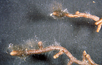
– Enlarged view – |
| • references | |
| Weiss M, Agerer R (1988) Studien an Ektomykorrhizen XII. Drei nichtidentifizierte Mykorrhizen an Picea abies (L.) Karst. aus einer Baumschule. Eur J For Path 18: 26-43. | |
| • ramification presence-type | |
| absent | |
| or | monopodial-pinnate |
| • ramification orders | |
| 0 | Lower value of unspecified range (could be µ-s.d., but not known) |
| 1 | Upper value of unspecified range (could be µ+s.d., but not known) |
| • abundance | |
| solitary or in small numbers | |
| • rhizomorphs as short mycorrhiza-like outgrowths with blunt tips presence | |
| absent | |
| • rhizomorphs presence | |
| absent | |
| • shape | |
| straight | |
| or | bent |
| • shape {of distal end} | |
| inflated, club-shaped | |
| • length | |
| 0.6 mm | Minimum value |
| 0.8 mm | Lower value of unspecified range (could be µ-s.d., but not known) |
| 1 mm | Upper value of unspecified range (could be µ+s.d., but not known) |
| • diameter | |
| 0.2 mm | Lower value of unspecified range (could be µ-s.d., but not known) |
| 0.3 mm | Upper value of unspecified range (could be µ+s.d., but not known) |
| • colour | |
| black | |
| • mantle transparency | |
| not transparent | |
| • mantle laticifers visibility | |
| absent | |
| • mantle dots presence-colour | |
| absent | |
| • mantle carbonizing presence | |
| absent | |
| • mantle surface {in general} habit | |
| smooth | |
| • mantle surface {in detail} kind | |
| loosely woolly | |
| • emanating hyphae distribution | |
| not specifically distributed | |
| • presence | |
| absent | |
| • blue granules presence | |
| absent | |
| • presence | |
| absent | |
| • organisation | |
| plectenchymatous | |
| • mantle type | |
| ring-like arrangement of hyphal bundles (type A) | |
| and | hyphae rather irregularly arranged and no special pattern discernible (type B) |
| • hyphal system kind | |
| undifferentiated | |
| and | forked |
| and | with rather short, obtuse, even finger-like branches |
| • hyphae hyphal junctions angle {between} | |
| ca. 45° and less | |
| • septa clamps presence | |
| absent | |
| • cell shape | |
| cylindric, constricted at septa | |
| and | slightly inflated in middle portions |
| • cell pigment location-colour | |
| membranaceously brownish | |
| • drops of exuded pigment presence | |
| absent | |
| • hyphae arrangement | |
| plectenchymatous, without pattern | |
| • organisation | |
| plectenchymatous | |
| • hyphae arrangement | |
| without pattern | |
| or | with some considerably inflated cells |
| • septa clamps presence | |
| absent | |
| • cell pigment location-colour | |
| absent | |
| • cell diameter | |
| 1 µm | Lower value of unspecified range (could be µ-s.d., but not known) |
| 6 µm | Upper value of unspecified range (could be µ+s.d., but not known) |
| • mantle thickness {apart from tip} | |
| 10 µm | Lower value of unspecified range (could be µ-s.d., but not known) |
| 20 µm | Upper value of unspecified range (could be µ+s.d., but not known) |
| • presence | |
| present | |
| • rows number | |
| 1 | Lower value of unspecified range (could be µ-s.d., but not known) |
| 3 | Upper value of unspecified range (could be µ+s.d., but not known) |
| • shape | |
| tangentially-oval, -elliptic or -cylindrical, and oriented in parallel to root axis | |
| • tangentially length | |
| 70 µm | Lower value of unspecified range (could be µ-s.d., but not known) |
| 110 µm | Upper value of unspecified range (could be µ+s.d., but not known) |
| • radially diameter | |
| 5 µm | Lower value of unspecified range (could be µ-s.d., but not known) |
| 10 µm | Upper value of unspecified range (could be µ+s.d., but not known) |
| • anatomy mantle longitudinal section cortical (epidermal) cells tangentially length | |
| 60 µm | Lower value of unspecified range (could be µ-s.d., but not known) |
| 90 µm | Upper value of unspecified range (could be µ+s.d., but not known) |
| • anatomy mantle longitudinal section cortical (epidermal) cells radially diameter | |
| 18 µm | Lower value of unspecified range (could be µ-s.d., but not known) |
| 30 µm | Upper value of unspecified range (could be µ+s.d., but not known) |
| • presence | |
| present | |
| • kind | |
| protruding towards endodermis | |
| • anatomy mantle longitudinal section hyphal cells around tannin cells shape | |
| roundish | |
| or | cylindrical |
| • anatomy mantle longitudinal section hyphal cells around tannin cells thickness | |
| 1.5 µm | Minimum value |
| 2 µm | Lower value of unspecified range (could be µ-s.d., but not known) |
| 3 µm | Upper value of unspecified range (could be µ+s.d., but not known) |
| 4 µm | Maximum value |
| • anatomy mantle longitudinal section hyphal rows around tannin cells number | |
| one | |
| • anatomy mantle longitudinal section hyphal cells around cortical (epidermal) cells shape | |
| roundish | |
| • anatomy mantle longitudinal section hyphal cells around cortical cells (epidermal) thickness | |
| 1.5 µm | Minimum value |
| 2 µm | Lower value of unspecified range (could be µ-s.d., but not known) |
| 3 µm | Upper value of unspecified range (could be µ+s.d., but not known) |
| 4 µm | Maximum value |
| • anatomy mantle longitudinal section hyphal rows around cortical (epidermal) cells number | |
| one | |
| • structure {in plan view} | |
| of palmetti type | |
| • lobes width | |
| 1 µm | Lower value of unspecified range (could be µ-s.d., but not known) |
| 3 µm | Upper value of unspecified range (could be µ+s.d., but not known) |
| • presence | |
| present | |
| • tangentially length | |
| 5 µm | Minimum value |
| 15 µm | Lower value of unspecified range (could be µ-s.d., but not known) |
| 25 µm | Upper value of unspecified range (could be µ+s.d., but not known) |
| • anatomy mantle cross-section cortical (epidermal) cells shape | |
| tangentially-oval to tangentially-elliptic | |
| • anatomy mantle cross-section cortical (epidermal) cells tangentially length | |
| 20 µm | Lower value of unspecified range (could be µ-s.d., but not known) |
| 30 µm | Upper value of unspecified range (could be µ+s.d., but not known) |
| • anatomy mantle cross-section hyphal cells around tannin cells shape | |
| roundish | |
| • anatomy mantle cross-section hyphal cells around tannin cells thickness | |
| 1.5 µm | Minimum value |
| 2 µm | Lower value of unspecified range (could be µ-s.d., but not known) |
| 3 µm | Upper value of unspecified range (could be µ+s.d., but not known) |
| 4 µm | Maximum value |
| • anatomy mantle cross-section hyphal rows around tannin cells number | |
| one | |
| or | two |
| • anatomy mantle cross-section hyphal cells around cortical (epidermal) cells thickness | |
| 1.5 µm | Minimum value |
| 2 µm | Lower value of unspecified range (could be µ-s.d., but not known) |
| 3 µm | Upper value of unspecified range (could be µ+s.d., but not known) |
| 4 µm | Maximum value |
| • anatomy mantle cross-section hyphal rows around cortical (epidermal) cells number | |
| one | |
| • intrahyphal hyphae presence | |
| absent | |
| • anastomoses type | |
| open, with a long bridge | |
| or | open, with a short bridge or bridge almost lacking |
| • shape | |
| not striking | |
| or | irregularly inflated or even beaded |
| • cell pigment location-colour | |
| membranaceously brownish | |
| • drops of exuded pigment presence | |
| absent | |
| • distance {from septum} | |
| in considerable distance from the septum | |
| • clamps presence | |
| absent | |
| • anatomy emanating elements emanating hyphae cell shape | |
| slightly constricted | |
| • anatomy emanating elements emanating hyphae cell diameter | |
| 1.5 µm | Minimum value |
| 2 µm | Mean (= average) |
| 3 µm | Maximum value |
| • anatomy emanating elements emanating hyphae cell length | |
| 18 µm | Minimum value |
| 30 µm | Lower value of unspecified range (could be µ-s.d., but not known) |
| 50 µm | Upper value of unspecified range (could be µ+s.d., but not known) |
| 60 µm | Maximum value |
| • anatomy emanating elements emanating hyphae cell wall surface habit | |
| smooth | |
| or | rough of warts |
| or | without lens-shaped appositions |
| or | without spindle-shaped appositions |
| • anatomy emanating elements emanating hyphae cell wall thickness | |
| 0.2 µm | Mean (= average) |
| • presence | |
| present | |
| • length | |
| 0 µm | Lower value of unspecified range (could be µ-s.d., but not known) |
| 0.5 µm | Upper value of unspecified range (could be µ+s.d., but not known) |
| 1 µm | Maximum value |
| • presence | |
| absent | |
| • {of ectomycorrhiza former} presence | |
| present | |
| • {of ectomycorrhiza former} abundance | |
| occasionally present | |
| • {in foreign ectomycorrhizae} presence | |
| absent | |
| • shape | |
| as normal hyphae | |
| • geographic occurrence continent | |
| Europe | |
| • plant family | |
| Pinaceae | |
| • plant genus | |
| Picea | |
| • plant habitat kind | |
| nursery | |
| • public notes | |
| Mycorrhizal systems monopodial, mostly simple. | |