|
|
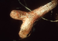
– Enlarged view – |
| • references | |
| Agerer R, ed (1987-1998) Colour Atlas of Ectomycorrhizae, 1st-11th delivery. Einhorn-Verlag, Schwäbisch Gmünd. Franz F (1994) Ektomykorrhizen der Fichte: Identifizierung, Ultrastruktur und Mikroelementanalyse (EELS, ESI). Diss Univ Bayreuth. Franz F, Acker G (1995) Rhizomorphs of Picea abies ectomycorrhizae: ultrastructural aspects and elemental analysis (EELS, ESI) on hyphal inclusions. Nova Hedwigia 60(1-2): 253-267. Haug I, Pritsch K (1992) Ectomycorrhizal tpyes of spruce (Picea abies (L.) Karst.) in the Black Forest. A microscopical atlas. Kernforschungszentrum Karlsruhe. Weiss M (1988) Ektomykorrhizen von Picea abies, Synthese, Ontogenie und Reaktion auf Umweltschadstoffe. Diss Univ München. |
|
| • length | |
| 0 mm | Lower value of unspecified range (could be µ-s.d., but not known) |
| 6 mm | Upper value of unspecified range (could be µ+s.d., but not known) |
| • ramification presence-type | |
| monopodial-pinnate | |
| or | monopodial-pyramidal |
| • rhizomorphs as stout, short, conical structures presence-abundance | |
| absent | |
| • exploration type | |
| medium distance smooth | |
| • shape | |
| straight | |
| or | bent |
| • shape {of distal end} | |
| not inflated, cylindric | |
| • length | |
| 0 mm | Lower value of unspecified range (could be µ-s.d., but not known) |
| 4 mm | Upper value of unspecified range (could be µ+s.d., but not known) |
| • diameter | |
| 0.3 mm | Lower value of unspecified range (could be µ-s.d., but not known) |
| 0.5 mm | Upper value of unspecified range (could be µ+s.d., but not known) |
| • colour | |
| red | |
| or | white |
| • older parts colour | |
| ochre, yellowish brown | |
| • mantle cortical cells visibility | |
| not visible | |
| • mantle {distinct} surface visibility | |
| present | |
| • mantle transparency | |
| not transparent | |
| • mantle laticifers visibility | |
| absent | |
| • mantle surface {in general} habit | |
| silvery | |
| or | smooth |
| or | not smooth |
| • mantle surface {in detail} kind | |
| densely cottony | |
| or | forming rings (reticulate) |
| • emanating hyphae presence | |
| present | |
| • emanating hyphae abundance | |
| abundant | |
| • diameter | |
| 0 mm | Lower value of unspecified range (could be µ-s.d., but not known) |
| 0.35 mm | Upper value of unspecified range (could be µ+s.d., but not known) |
| • cross-section shape | |
| round or roundish | |
| • colour | |
| concolourous to mantle | |
| or | red |
| or | whitish |
| • ramification kind-frequency | |
| repeatedly into smaller filaments | |
| • connection to mantle kind | |
| distinct | |
| • margin habit | |
| hairy | |
| • dimorphism presence | |
| absent | |
| • presence | |
| absent | |
| • emanating elements presence-type | |
| rhizomorphs | |
| • presence | |
| absent | |
| • organisation | |
| plectenchymatous | |
| • mantle type | |
| ring-like arrangement of hyphal bundles (type A) | |
| • cell diameter | |
| 2 µm | Minimum value |
| 2.5 µm | Lower value of unspecified range (could be µ-s.d., but not known) |
| 4 µm | Upper value of unspecified range (could be µ+s.d., but not known) |
| 5 µm | Maximum value |
| • cell length | |
| 10 µm | Lower value of unspecified range (could be µ-s.d., but not known) |
| 90 µm | Upper value of unspecified range (could be µ+s.d., but not known) |
| • cell wall surface habit | |
| smooth | |
| and | with few crystals |
| • organisation | |
| plectenchymatous | |
| • hyphae arrangement | |
| plectenchymatous, ring-like | |
| • cell pigment location-colour | |
| colourless | |
| • cell diameter | |
| 2 µm | Minimum value |
| 3 µm | Lower value of unspecified range (could be µ-s.d., but not known) |
| 6 µm | Upper value of unspecified range (could be µ+s.d., but not known) |
| • organisation | |
| plectenchymatous | |
| • hyphae arrangement | |
| ring-like | |
| • mantle thickness {apart from tip} | |
| 15 µm | Lower value of unspecified range (could be µ-s.d., but not known) |
| 30 µm | Upper value of unspecified range (could be µ+s.d., but not known) |
| • mantle different layers presence | |
| not discernable | |
| • outer mantle layer organisation | |
| plectenchymatous | |
| • middle mantle layer organisation | |
| plectenchymatous | |
| • inner mantle layer organisation | |
| plectenchymatous | |
| • inner mantle layer hyphae radially diameter | |
| 2 µm | Lower value of unspecified range (could be µ-s.d., but not known) |
| 6 µm | Upper value of unspecified range (could be µ+s.d., but not known) |
| • presence | |
| present | |
| • presence | |
| present | |
| • kind | |
| protruding towards endodermis | |
| • anatomy mantle longitudinal section hyphal cells around tannin cells thickness | |
| 3 µm | Lower value of unspecified range (could be µ-s.d., but not known) |
| 6 µm | Upper value of unspecified range (could be µ+s.d., but not known) |
| • anatomy mantle longitudinal section hyphal cells around cortical cells (epidermal) thickness | |
| 2.5 µm | Lower value of unspecified range (could be µ-s.d., but not known) |
| 4 µm | Upper value of unspecified range (could be µ+s.d., but not known) |
| • structure {in plan view} | |
| of palmetti type | |
| • lobes width | |
| 1 µm | Minimum value |
| 2 µm | Lower value of unspecified range (could be µ-s.d., but not known) |
| 3 µm | Upper value of unspecified range (could be µ+s.d., but not known) |
| 5 µm | Maximum value |
| • intrahyphal hyphae presence | |
| present | |
| • septal pores configuration | |
| dolipore-like structures | |
| • shape | |
| not striking | |
| • clamps presence | |
| present | |
| • presence | |
| absent | |
| or | present |
| • abundance | |
| infrequent | |
| • distribution | |
| not specified | |
| • anatomy emanating elements emanating hyphae cell diameter | |
| 2 µm | Lower value of unspecified range (could be µ-s.d., but not known) |
| 4 µm | Upper value of unspecified range (could be µ+s.d., but not known) |
| • anatomy emanating elements emanating hyphae cell wall surface habit | |
| smooth | |
| • type | |
| undifferentiated; hyphae rather loosely woven and of uniform diameter (type A) |
|
| • presence | |
| absent | |
| • {of ectomycorrhiza former} presence | |
| absent | |
| • septal pores type | |
| as dolipores with perforated parenthesome | |
| • reaction with KOH 10% presence | |
| present | |
| • geographic occurrence continent | |
| Europe | |
| • plant family | |
| Pinaceae | |
| • plant genus | |
| Picea | |
| • plant habitat kind | |
| forests, woods | |
| • family | |
| Thelephoraceae ss. Stalpers. excl. Tylospora | |
| • public notes | |
| Mycorrhizal ends somtimes sightly bent, whitish rose-red, in older parts brownish, rhizomorphs rose-red to white; emanating hyphae septate with clamps (? Haug & Pritsch 1992), epimembranaceously reddish; mantle in KOH intensely violet. | |