 |
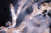
– Enlarged view – |
| • references | |
| Ingleby K, Mason PA, Last FT, Fleming LV (1990) Identification of ectomycorrhizas. ITE Research Publication no. 5. HMSO, London. | |
| • length | |
| 0 mm | Lower value of unspecified range (could be µ-s.d., but not known) |
| 5 mm | Upper value of unspecified range (could be µ+s.d., but not known) |
| • ramification presence-type | |
| monopodial-pinnate | |
| • ramification orders | |
| 0 | Lower value of unspecified range (could be µ-s.d., but not known) |
| 1 | Upper value of unspecified range (could be µ+s.d., but not known) |
| • main axis diameter | |
| 0 mm | Lower value of unspecified range (could be µ-s.d., but not known) |
| 3 mm | Upper value of unspecified range (could be µ+s.d., but not known) |
| • rhizomorphs as stout, short, conical structures presence-abundance | |
| absent | |
| • rhizomorphs as short mycorrhiza-like outgrowths with blunt tips presence | |
| absent | |
| • rhizomorphs presence | |
| absent | |
| • shape | |
| bent | |
| • shape {of distal end} | |
| not inflated, cylindric | |
| • colour | |
| white | |
| • very tip colour | |
| white | |
| • older parts colour | |
| brown | |
| or | grey |
| • mantle transparency | |
| not transparent | |
| • mantle laticifers visibility | |
| absent | |
| • mantle dots presence-colour | |
| absent | |
| • mantle carbonizing presence | |
| absent | |
| • mantle surface {in general} habit | |
| silvery | |
| • mantle surface {in detail} kind | |
| densely cottony | |
| or | loosely cottony |
| • emanating hyphae presence | |
| present | |
| • emanating hyphae abundance | |
| abundant | |
| • presence | |
| present | |
| • abundance | |
| infrequent | |
| • shape | |
| globular | |
| • diameter | |
| 0.2 mm | Lower value of unspecified range (could be µ-s.d., but not known) |
| 0.4 mm | Upper value of unspecified range (could be µ+s.d., but not known) |
| • colour | |
| white | |
| • formation location | |
| on the mantle | |
| • presence | |
| absent | |
| • organisation | |
| plectenchymatous | |
| • mantle type | |
| hyphae rather irregularly arranged and no special pattern discernible (type B) | |
| • hyphal system kind | |
| undifferentiated | |
| • septa clamps presence | |
| absent | |
| • cell wall surface habit | |
| smooth | |
| and | rough |
| • drops of exuded pigment presence | |
| absent | |
| • hyphae arrangement | |
| plectenchymatous, without pattern | |
| • organisation | |
| plectenchymatous with pseudoparenchymatous nests of cells | |
| • hyphae arrangement | |
| ring-like | |
| • cell diameter | |
| 0 µm | Lower value of unspecified range (could be µ-s.d., but not known) |
| 7 µm | Upper value of unspecified range (could be µ+s.d., but not known) |
| • cell length | |
| 0 µm | Lower value of unspecified range (could be µ-s.d., but not known) |
| 80 µm | Upper value of unspecified range (could be µ+s.d., but not known) |
| • mantle thickness {apart from tip} | |
| 5 µm | Lower value of unspecified range (could be µ-s.d., but not known) |
| 15 µm | Upper value of unspecified range (could be µ+s.d., but not known) |
| • anastomoses anastomosal bridge thickness {relative to hyphae} | |
| as thick as | |
| • shape | |
| not striking | |
| • drops of exuded pigment presence | |
| absent | |
| • presence | |
| present | |
| • kind | |
| acute | |
| • side-branches at septum number | |
| one side-branch at septum | |
| • clamps presence | |
| present | |
| • clamps hole presence | |
| absent | |
| • presence | |
| present | |
| • abundance | |
| infrequent | |
| • distribution | |
| not specified | |
| • anatomy emanating elements emanating hyphae cell diameter | |
| 2 µm | Lower value of unspecified range (could be µ-s.d., but not known) |
| 4 µm | Upper value of unspecified range (could be µ+s.d., but not known) |
| • anatomy emanating elements emanating hyphae cell wall surface habit | |
| smooth | |
| or | rough of warts |
| • type | |
| lacking, only emanating hyphae present (type G) |
|
| • internal structure organisation | |
| pseudoparenchymatous with angular cells | |
| or | pseudoparenchymatous with roundish cells |
| or | outer layer plectenchymatous, center pseudoparenchymatous |
| • center structure habit | |
| compactly filled | |
| • geographic occurrence continent | |
| Europe | |
| • plant family | |
| Pinaceae | |
| • plant genus | |
| Picea | |
| • plant habitat kind | |
| nursery | |
| • family | |
| Cortinariaceae | |
| • fruitbodies growth habit | |
| epigeous | |
| or | pileate-lamellate |
| • public notes | |
| Mycorrhizal systems infrequently branched; mycorrhizal ends short, slender, silver-white when young, older parts changing to grey-brown. | |