|
|
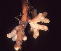
– Enlarged view – |
| • references | |
| Treu R (1990) Charakterisierung und Identifizierung von Ektomykorrhizen aus dem Nationalpark Berchtesgaden. Bibl Mycol 134: 1-196. Treu R (1990) Russula firmula. In Agerer R (ed) Colour Atlas of Ectomycorrhizae, plate 43, Einhorn-Verlag, Schwäbisch Gmünd. |
|
| • length | |
| 0 mm | Lower value of unspecified range (could be µ-s.d., but not known) |
| 6 mm | Upper value of unspecified range (could be µ+s.d., but not known) |
| • ramification presence-type | |
| dichotomous | |
| • main axis diameter | |
| 0.4 mm | Lower value of unspecified range (could be µ-s.d., but not known) |
| 0.53 mm | Upper value of unspecified range (could be µ+s.d., but not known) |
| • rhizomorphs as stout, short, conical structures presence-abundance | |
| absent | |
| • rhizomorphs as short mycorrhiza-like outgrowths with blunt tips presence | |
| absent | |
| • rhizomorphs presence | |
| absent | |
| • shape | |
| straight | |
| • shape {of distal end} | |
| not inflated, cylindric | |
| • length | |
| 0 mm | Lower value of unspecified range (could be µ-s.d., but not known) |
| 2.3 mm | Upper value of unspecified range (could be µ+s.d., but not known) |
| • diameter | |
| 0.33 mm | Lower value of unspecified range (could be µ-s.d., but not known) |
| 0.5 mm | Upper value of unspecified range (could be µ+s.d., but not known) |
| • colour | |
| yellow | |
| or | orange |
| • very tip colour | |
| yellowish | |
| • older parts colour | |
| brown | |
| or | red |
| • mantle cortical cells visibility | |
| not visible | |
| • mantle {distinct} surface visibility | |
| present | |
| • mantle transparency | |
| not transparent | |
| • mantle laticifers visibility | |
| absent | |
| • mantle dots presence-colour | |
| absent | |
| • mantle carbonizing presence | |
| absent | |
| • mantle surface {in general} habit | |
| smooth | |
| • presence | |
| absent | |
| • blue granules presence | |
| absent | |
| • presence | |
| absent | |
| • anatomy entire mycorrhiza laticiferous solitary (gloeoplerous) cells presence | |
| present | |
| • organisation | |
| pseudoparenchymatous | |
| • organisation {if pseudoparenchymatous} cell shape | |
| angular | |
| • mantle type | |
| some cells containing droplets, staining in sulpho-vanillin; shape variable (type N) | |
| and | angular cells bearing a hyphal net (type P) |
| • septa clamps presence | |
| absent | |
| • cell diameter | |
| 3 µm | Minimum value |
| 6 µm | Lower value of unspecified range (could be µ-s.d., but not known) |
| 17 µm | Upper value of unspecified range (could be µ+s.d., but not known) |
| • cell length | |
| 3 µm | Minimum value |
| 9 µm | Lower value of unspecified range (could be µ-s.d., but not known) |
| 22 µm | Upper value of unspecified range (could be µ+s.d., but not known) |
| • cell wall thickness | |
| 0.2 µm | Lower value of unspecified range (could be µ-s.d., but not known) |
| 0.5 µm | Upper value of unspecified range (could be µ+s.d., but not known) |
| 1 µm | Maximum value |
| • cell wall surface habit | |
| smooth | |
| • drops of exuded pigment presence | |
| absent | |
| • organisation | |
| pseudoparenchymatous | |
| • cell diameter | |
| 4 µm | Lower value of unspecified range (could be µ-s.d., but not known) |
| 12 µm | Upper value of unspecified range (could be µ+s.d., but not known) |
| • cell length | |
| 1 µm | Mean (= average) |
| • cell density | |
| 8 | Lower value of unspecified range (could be µ-s.d., but not known) |
| 12 | Upper value of unspecified range (could be µ+s.d., but not known) |
| • cell wall thickness | |
| 0.2 µm | Mean (= average) |
| • cell wall surface habit | |
| smooth | |
| • organisation | |
| plectenchymatous | |
| • hyphae arrangement | |
| star-like | |
| • septa clamps presence | |
| absent | |
| • cell diameter | |
| 2 µm | Lower value of unspecified range (could be µ-s.d., but not known) |
| 4 µm | Upper value of unspecified range (could be µ+s.d., but not known) |
| • anatomy mantle outer mantle layer {of ectomycorrhizal tip} organisation | |
| like other parts of mantle | |
| • mantle thickness {apart from tip} | |
| 15 µm | Lower value of unspecified range (could be µ-s.d., but not known) |
| 20 µm | Upper value of unspecified range (could be µ+s.d., but not known) |
| • mantle thickness {at ectomycorrhizal tip} | |
| 20 µm | Mean (= average) |
| • mantle different layers presence | |
| not discernable | |
| • outer mantle layer organisation | |
| pseudoparenchymatous | |
| • middle mantle layer organisation | |
| pseudoparenchymatous | |
| • inner mantle layer organisation | |
| pseudoparenchymatous | |
| • unlayered mantle hyphae tangentially length | |
| 3 µm | Lower value of unspecified range (could be µ-s.d., but not known) |
| 15 µm | Upper value of unspecified range (could be µ+s.d., but not known) |
| • unlayered mantle hyphae radially diameter | |
| 2 µm | Lower value of unspecified range (could be µ-s.d., but not known) |
| 5 µm | Upper value of unspecified range (could be µ+s.d., but not known) |
| • anatomy mantle longitudinal section cortical (epidermal) cells shape | |
| radially-oval to -elliptic | |
| or | tangentially-oval to -elliptic or -cylindrical, and oriented in parallel to root axis |
| • anatomy mantle longitudinal section cortical (epidermal) cells tangentially length | |
| 15 µm | Lower value of unspecified range (could be µ-s.d., but not known) |
| 43 µm | Upper value of unspecified range (could be µ+s.d., but not known) |
| • anatomy mantle longitudinal section cortical (epidermal) cells radially diameter | |
| 15 µm | Lower value of unspecified range (could be µ-s.d., but not known) |
| 53 µm | Upper value of unspecified range (could be µ+s.d., but not known) |
| • anatomy mantle longitudinal section cortical (epidermal) cells mean tangential length CCt (ECt) | |
| 29.6 µm | Mean (= average) |
| • anatomy mantle longitudinal section cortical (epidermal) cells mean shape-ratio CCq (ECq) | |
| 1.1 | Mean (= average) |
| • mantle different layers presence | |
| not discernible | |
| • outer mantle layer organisation | |
| pseudoparenchymatous | |
| • middle mantle layer organisation | |
| pseudoparenchymatous | |
| • inner mantle layer organisation | |
| pseudoparenchymatous | |
| • unlayered mantle hyphae tangentially length | |
| 4 µm | Lower value of unspecified range (could be µ-s.d., but not known) |
| 20 µm | Upper value of unspecified range (could be µ+s.d., but not known) |
| • unlayered mantle hyphae radially diameter | |
| 2 µm | Lower value of unspecified range (could be µ-s.d., but not known) |
| 5 µm | Upper value of unspecified range (could be µ+s.d., but not known) |
| • presence | |
| present | |
| • anatomy mantle cross-section cortical (epidermal) cells shape | |
| tangentially-oval to tangentially-elliptic | |
| • anatomy mantle cross-section cortical (epidermal) cells tangentially length | |
| 30 µm | Lower value of unspecified range (could be µ-s.d., but not known) |
| 90 µm | Upper value of unspecified range (could be µ+s.d., but not known) |
| • anatomy mantle cross-section cortical (epidermal) cells radially diameter | |
| 18 µm | Lower value of unspecified range (could be µ-s.d., but not known) |
| 40 µm | Upper value of unspecified range (could be µ+s.d., but not known) |
| • anatomy mantle cross-section cortical (epidermal) cells mean tangential length CCt | |
| 61.5 µm | Mean (= average) |
| • anatomy mantle cross-section cortical (epidermal) cells mean shape-ratio CCq | |
| 2.3 | Mean (= average) |
| • anatomy mantle cross-section hyphal cells around cortical (epidermal) cells shape | |
| cylindrical | |
| • anatomy mantle cross-section hyphal cells around cortical (epidermal) cells thickness | |
| 2 µm | Lower value of unspecified range (could be µ-s.d., but not known) |
| 3.5 µm | Upper value of unspecified range (could be µ+s.d., but not known) |
| • anatomy mantle cross-section hyphal rows around cortical (epidermal) cells number | |
| one | |
| • clamps presence | |
| absent | |
| • type | |
| lacking, only emanating hyphae present (type G) |
|
| • presence | |
| absent | |
| • {of ectomycorrhiza former} presence | |
| absent | |
| • {of foreign origin} presence | |
| absent | |
| • number {per cell} | |
| 1 | Minimum value |
| 2 | Mean (= average) |
| • shape | |
| round | |
| • diameter | |
| 1 µm | Lower value of unspecified range (could be µ-s.d., but not known) |
| 2.5 µm | Upper value of unspecified range (could be µ+s.d., but not known) |
| • length | |
| 1 µm | Lower value of unspecified range (could be µ-s.d., but not known) |
| 2.5 µm | Upper value of unspecified range (could be µ+s.d., but not known) |
| • geographic occurrence continent | |
| Europe | |
| • knowledge about association with foreign fruitbodies presence | |
| unknown | |
| • plant family | |
| Pinaceae | |
| • plant genus | |
| Pinus | |
| • plant subgenus-section | |
| two-needle-pines | |
| • plant habitat kind | |
| forests, woods | |
| • family | |
| Russulaceae | |
| • subgenus-section | |
| Russula sect. Russula | |
| • fruitbodies growth habit | |
| epigeous | |
| or | pileate-lamellate |
| • public notes | |
| Mycorrrhizal ends orange-yellow, older parts reddish brown; autofluorescence of mantle in section with UV-filter in outer parts yellowish in inner parts bluish, with blue-filter yellowish; mantle in sulfo-vanillin with some cells staining blue-black. | |