|
|
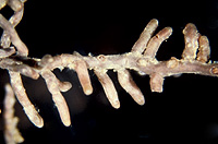
– Enlarged view – |
| • references | |
| Agerer R (1986) Studies on ectomycorrhizae III. Mycorrhizae formed by four fungi in the genera Lactarius and Russula on spruce. Mycotaxon 27: 1-59. Agerer R (1987) Russula xerampelina. In Agerer R (ed) Colour Atlas of Ectomycorrhizae, plate 2, Einhorn-Verlag, Schwäbisch Gmünd. - |
|
| • ramification presence-type | |
| monopodial-pinnate | |
| • tips {per 10 mm} number | |
| 0 | Lower value of unspecified range (could be µ-s.d., but not known) |
| 1 | Upper value of unspecified range (could be µ+s.d., but not known) |
| • rhizomorphs as stout, short, conical structures presence-abundance | |
| absent | |
| • rhizomorphs as short mycorrhiza-like outgrowths with blunt tips presence | |
| absent | |
| • rhizomorphs presence | |
| present | |
| • rhizomorphs frequency | |
| infrequent | |
| • exploration type | |
| contact | |
| • shape | |
| straight | |
| or | bent |
| • shape {of distal end} | |
| not inflated, cylindric | |
| • length | |
| 0 mm | Lower value of unspecified range (could be µ-s.d., but not known) |
| 2 mm | Upper value of unspecified range (could be µ+s.d., but not known) |
| • diameter | |
| 0.24 mm | Minimum value |
| 0.33 mm | Lower value of unspecified range (could be µ-s.d., but not known) |
| 0.39 mm | Upper value of unspecified range (could be µ+s.d., but not known) |
| 0.48 mm | Maximum value |
| • colour | |
| brown | |
| • older parts colour | |
| dark brown | |
| or | red |
| • mantle cortical cells visibility | |
| not visible | |
| • mantle {distinct} surface visibility | |
| present | |
| • mantle transparency | |
| not transparent | |
| • mantle laticifers visibility | |
| absent | |
| • mantle dots presence-colour | |
| absent | |
| • mantle carbonizing presence | |
| absent | |
| • mantle surface {in general} habit | |
| shiny | |
| or | smooth |
| • emanating hyphae presence | |
| present | |
| • emanating hyphae abundance | |
| infrequent | |
| • cross-section shape | |
| round or roundish | |
| • colour | |
| whitish | |
| • origin location | |
| proximal | |
| • dimorphism presence | |
| absent | |
| • presence | |
| absent | |
| • emanating elements presence-type | |
| rhizomorphs | |
| or | cystidia |
| • emanating elements cystidia location | |
| on outer mantle layer | |
| • presence | |
| absent | |
| • organisation | |
| plectenchymatous | |
| • mantle type | |
| a net of coarse and irregularly shaped hyphae (type H) | |
| • septa thickness {relative to cell walls} | |
| as thick as walls | |
| • septa clamps presence | |
| absent | |
| • cell shape | |
| cylindric, not constricted at septa | |
| • cell diameter | |
| 4 µm | Minimum value |
| 5 µm | Lower value of unspecified range (could be µ-s.d., but not known) |
| 7 µm | Upper value of unspecified range (could be µ+s.d., but not known) |
| • cell wall thickness | |
| 0.2 µm | Lower value of unspecified range (could be µ-s.d., but not known) |
| 1 µm | Upper value of unspecified range (could be µ+s.d., but not known) |
| • cell wall surface habit | |
| smooth | |
| • drops of exuded pigment presence | |
| absent | |
| • organisation | |
| plectenchymatous | |
| • hyphae arrangement | |
| without pattern | |
| • septa clamps presence | |
| absent | |
| • cell diameter | |
| 2.5 µm | Lower value of unspecified range (could be µ-s.d., but not known) |
| 3 µm | Upper value of unspecified range (could be µ+s.d., but not known) |
| • anatomy mantle outer mantle layer {of ectomycorrhizal tip} organisation | |
| like other parts of mantle | |
| or | plectenchymatous |
| • mantle thickness {apart from tip} | |
| 20 µm | Lower value of unspecified range (could be µ-s.d., but not known) |
| 30 µm | Upper value of unspecified range (could be µ+s.d., but not known) |
| • mantle different layers presence | |
| discernable | |
| • outer mantle layer organisation | |
| pseudoparenchymatous | |
| • outer mantle layer hyphae tangentially length | |
| 6 µm | Lower value of unspecified range (could be µ-s.d., but not known) |
| 10 µm | Upper value of unspecified range (could be µ+s.d., but not known) |
| 15 µm | Maximum value |
| • outer mantle layer hyphae radially diameter | |
| 2 µm | Lower value of unspecified range (could be µ-s.d., but not known) |
| 4 µm | Upper value of unspecified range (could be µ+s.d., but not known) |
| • middle mantle layer organisation | |
| pseudoparenchymatous | |
| • middle mantle layer hyphae tangentially length | |
| 5 µm | Lower value of unspecified range (could be µ-s.d., but not known) |
| 15 µm | Upper value of unspecified range (could be µ+s.d., but not known) |
| • middle mantle layer hyphae radially diameter | |
| 3 µm | Lower value of unspecified range (could be µ-s.d., but not known) |
| 6 µm | Upper value of unspecified range (could be µ+s.d., but not known) |
| 8 µm | Maximum value |
| • inner mantle layer organisation | |
| pseudoparenchymatous | |
| • inner mantle layer hyphae tangentially length | |
| 3 µm | Lower value of unspecified range (could be µ-s.d., but not known) |
| 8 µm | Upper value of unspecified range (could be µ+s.d., but not known) |
| • inner mantle layer hyphae radially diameter | |
| 2 µm | Lower value of unspecified range (could be µ-s.d., but not known) |
| 3 µm | Upper value of unspecified range (could be µ+s.d., but not known) |
| • presence | |
| present | |
| • shape | |
| tangentially-oval, -elliptic or -cylindrical, and oriented in parallel to root axis | |
| • tangentially length | |
| 30 µm | Lower value of unspecified range (could be µ-s.d., but not known) |
| 60 µm | Upper value of unspecified range (could be µ+s.d., but not known) |
| 80 µm | Maximum value |
| • radially diameter | |
| 4 µm | Lower value of unspecified range (could be µ-s.d., but not known) |
| 10 µm | Upper value of unspecified range (could be µ+s.d., but not known) |
| • anatomy mantle longitudinal section cortical (epidermal) cells shape | |
| tangentially-oval to -elliptic or -cylindrical, and oriented obliquely | |
| • anatomy mantle longitudinal section cortical (epidermal) cells tangentially length | |
| 30 µm | Minimum value |
| 40 µm | Lower value of unspecified range (could be µ-s.d., but not known) |
| 60 µm | Upper value of unspecified range (could be µ+s.d., but not known) |
| 80 µm | Maximum value |
| • anatomy mantle longitudinal section cortical (epidermal) cells radially diameter | |
| 12 µm | Minimum value |
| 15 µm | Lower value of unspecified range (could be µ-s.d., but not known) |
| 22 µm | Upper value of unspecified range (could be µ+s.d., but not known) |
| 28 µm | Maximum value |
| • mantle different layers presence | |
| discernible | |
| • outer mantle layer organisation | |
| pseudoparenchymatous | |
| • outer mantle layer hyphae tangentially length | |
| 2 µm | Minimum value |
| 6 µm | Lower value of unspecified range (could be µ-s.d., but not known) |
| 10 µm | Upper value of unspecified range (could be µ+s.d., but not known) |
| 14 µm | Maximum value |
| • outer mantle layer hyphae radially diameter | |
| 2 µm | Lower value of unspecified range (could be µ-s.d., but not known) |
| 2.5 µm | Upper value of unspecified range (could be µ+s.d., but not known) |
| • middle mantle layer organisation | |
| pseudoparenchymatous | |
| • middle mantle layer hyphae tangentially length | |
| 2 µm | Minimum value |
| 5 µm | Lower value of unspecified range (could be µ-s.d., but not known) |
| 9 µm | Upper value of unspecified range (could be µ+s.d., but not known) |
| 15 µm | Maximum value |
| • middle mantle layer hyphae radially diameter | |
| 2 µm | Minimum value |
| 3 µm | Lower value of unspecified range (could be µ-s.d., but not known) |
| 4 µm | Upper value of unspecified range (could be µ+s.d., but not known) |
| 5 µm | Maximum value |
| • inner mantle layer organisation | |
| pseudoparenchymatous | |
| • inner mantle layer hyphae tangentially length | |
| 2 µm | Lower value of unspecified range (could be µ-s.d., but not known) |
| 3 µm | Upper value of unspecified range (could be µ+s.d., but not known) |
| • inner mantle layer hyphae radially diameter | |
| 2 µm | Lower value of unspecified range (could be µ-s.d., but not known) |
| 3 µm | Upper value of unspecified range (could be µ+s.d., but not known) |
| • presence | |
| present | |
| • rows number | |
| 2 | Mean (= average) |
| 3 | Maximum value |
| • shape | |
| tangentially-oval to tangentially-elliptic | |
| • tangentially length | |
| 50 µm | Lower value of unspecified range (could be µ-s.d., but not known) |
| 65 µm | Upper value of unspecified range (could be µ+s.d., but not known) |
| • radially diameter | |
| 2 µm | Lower value of unspecified range (could be µ-s.d., but not known) |
| 17 µm | Upper value of unspecified range (could be µ+s.d., but not known) |
| 20 µm | Maximum value |
| • anatomy mantle cross-section cortical (epidermal) cells shape | |
| radially-oval to -elliptic | |
| • anatomy mantle cross-section cortical (epidermal) cells tangentially length | |
| 10 µm | Lower value of unspecified range (could be µ-s.d., but not known) |
| 20 µm | Upper value of unspecified range (could be µ+s.d., but not known) |
| • anatomy mantle cross-section cortical (epidermal) cells radially diameter | |
| 20 µm | Minimum value |
| 27 µm | Lower value of unspecified range (could be µ-s.d., but not known) |
| 35 µm | Upper value of unspecified range (could be µ+s.d., but not known) |
| • presence | |
| present | |
| • kind | |
| protruding towards endodermis | |
| • anatomy mantle cross-section hyphal cells around tannin cells shape | |
| roundish | |
| • anatomy mantle cross-section hyphal rows around tannin cells number | |
| one | |
| or | two |
| or | three |
| • anatomy mantle cross-section hyphal cells around cortical (epidermal) cells shape | |
| roundish | |
| • anatomy mantle cross-section hyphal rows around cortical (epidermal) cells number | |
| one | |
| • backwards-oriented clamps presence | |
| absent | |
| • anastomoses location | |
| not specified | |
| • type | |
| flask-shaped with an apical knob (type D) | |
| • apical knobs number | |
| 1 | Mean (= average) |
| • diameter {proximal} | |
| 5 µm | Lower value of unspecified range (could be µ-s.d., but not known) |
| 6 µm | Upper value of unspecified range (could be µ+s.d., but not known) |
| • length | |
| 30 µm | Lower value of unspecified range (could be µ-s.d., but not known) |
| 40 µm | Upper value of unspecified range (could be µ+s.d., but not known) |
| • shape | |
| not striking | |
| • cell pigment location-colour | |
| absent | |
| • drops of exuded pigment presence | |
| absent | |
| • clamps presence | |
| absent | |
| • clamps hole presence | |
| absent | |
| • clamps blister-like structure {at basis} presence | |
| absent | |
| • anatomy emanating elements emanating hyphae cell shape | |
| even | |
| • anatomy emanating elements emanating hyphae cell diameter | |
| 1.5 µm | Lower value of unspecified range (could be µ-s.d., but not known) |
| 2 µm | Upper value of unspecified range (could be µ+s.d., but not known) |
| • anatomy emanating elements emanating hyphae cell wall surface habit | |
| smooth | |
| or | without lens-shaped appositions |
| or | without spindle-shaped appositions |
| • anatomy emanating elements emanating hyphae cell wall thickness | |
| 0.2 µm | Mean (= average) |
| • anatomy emanating elements emanating hyphae cell wall thickness at tip {relative to remaining cell wall} | |
| thinner | |
| • anatomy emanating elements emanating hyphae cell wall {apart from tip} evenness | |
| even in thickness | |
| • type | |
| differentiated; thick hyphae forming a core, septa complete or sometimes enlarged (type E) |
|
| • internal nodia presence | |
| absent | |
| • cup-like structures on surface presence | |
| absent | |
| • a "ball" of intertwined, ramified, thin hyphae presence | |
| absent | |
| • ampullate, trumpet-like inflated presence | |
| absent | |
| • anatomy emanating elements rhizomorphs hyphae ampullate, trumpet-like inflated presence | |
| absent | |
| • anatomy emanating elements rhizomorphs hyphae ampullate, trumpet-like inflated presence | |
| absent | |
| • anatomy emanating elements rhizomorphs hyphae ampullate, trumpet-like inflated presence | |
| absent | |
| • anatomy emanating elements rhizomorphs hyphae ampullate, trumpet-like inflated presence | |
| absent | |
| • presence | |
| absent | |
| • {of ectomycorrhiza former} presence | |
| absent | |
| • number {per cell} | |
| 2 | Mean (= average) |
| • presence | |
| present | |
| • frequency | |
| in few cells | |
| • geographic occurrence continent | |
| Europe | |
| • plant family | |
| Pinaceae | |
| • plant genus | |
| Picea | |
| • plant habitat kind | |
| forests, woods | |
| • family | |
| Russulaceae | |
| • subgenus-section | |
| Russula sect. Rigidae | |
| • subsection | |
| Russula sect. Rigidae subsect. Xerampelinae | |
| • fruitbodies growth habit | |
| epigeous | |
| or | pileate-lamellate |
| • public notes | |
| Mycorrhizal ends more or less straight, dark flesh-brown, older parts dark reddish brown; autofluorescence of mantle in section with UV-filter slightly blue, with blue-filter greenish yellow; mantle hyphae in sulfo-vanillin pinkish, in brillant-cresyl-blue bright blue and after treatment with water violet-blue, in toluidin-blue violet-blue, in cotton-blue walls distinctly blue, in acid fuchsin slightly reddish; nuclei lying close together. | |