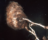
|
|
– Enlarged view – |
| • references | |
| Agerer R (1990) Studies on ectomycorrhizae XXIV. Ectomycorrhizae of Chroogomphus helveticus and C. rutilus (Gomphidiaceae, Basidiomycetes) and their rela- tionship to those of Suillus and Rhizopogon. Nova Hedwigia 50(1-2): 1-63. Treu R (1990) Charakterisierung und Identifizierung von Ektomykorrhizen aus den Nationalpark Berchtesgaden. Bibl Mycol 134: 1-196. Treu R (1990) Suillus plorans. In Agerer R (ed) Colour Atlas of Ectomycorrhizae, plate 46, Einhorn-Verlag, Schwäbisch Gmünd. Mleczko P, Ronikier M (2007) Features of ectomycorrhizae confirm molecular phylogenetics of Suillus (Boletales) rather than carpophore-based systematics: insights from studies on Suillus variegatus, S. plorans and related species. Nova Hedwegia 84(1-2): 1-20. |
|
| • length | |
| 0 mm | Lower value of unspecified range (could be µ-s.d., but not known) |
| 10 mm | Upper value of unspecified range (could be µ+s.d., but not known) |
| 20 mm | Maximum value |
| • ramification presence-type | |
| tubercle-like | |
| • surface {if tubercle-like} | |
| stringy | |
| or | woolly |
| • colour {if tubercle-like} | |
| ochre, yellowish brown [older reddish-brown] | |
| or | whitish [covered with pinkish or reddish brown dots and veins] |
| • rhizomorphs as stout, short, conical structures presence-abundance | |
| absent | |
| • rhizomorphs as short mycorrhiza-like outgrowths with blunt tips presence | |
| absent | |
| • rhizomorphs presence | |
| present | |
| • rhizomorphs frequency | |
| infrequent | |
| or | abundant [common] |
| • exploration type | |
| long distance | |
| • shape | |
| straight | |
| or | bent |
| • shape {of distal end} | |
| not inflated, cylindric | |
| • length | |
| 0 mm | Lower value of unspecified range (could be µ-s.d., but not known) |
| 2 mm | Upper value of unspecified range (could be µ+s.d., but not known) |
| • colour | |
| ochre, yellowish brown | |
| or | white |
| or | whitish |
| • very tip colour | |
| brown | |
| or | reddish |
| • mantle cortical cells visibility | |
| not visible | |
| • mantle {distinct} surface visibility | |
| present | |
| • mantle transparency | |
| not transparent | |
| • mantle laticifers visibility | |
| absent | |
| • mantle dots presence-colour | |
| absent | |
| • mantle carbonizing presence | |
| absent | |
| • mantle surface {in general} habit | |
| silvery | |
| • diameter | |
| 0 mm | Lower value of unspecified range (could be µ-s.d., but not known) |
| 0.64 mm | Upper value of unspecified range (could be µ+s.d., but not known) |
| • cross-section shape | |
| round or roundish | |
| • ramification kind-frequency | |
| infrequently, at restricted points | |
| • connection to mantle kind | |
| distinct | |
| • origin location | |
| proximal | |
| • margin habit | |
| hairy | |
| • dimorphism presence | |
| absent | |
| • presence | |
| absent | |
| • emanating elements presence-type | |
| rhizomorphs | |
| • blue granules presence | |
| absent | |
| • presence | |
| absent | |
| • matrix presence | |
| present | |
| • matrix location | |
| outer mantle layer {apart from tip} | |
| • organisation | |
| plectenchymatous | |
| • mantle type | |
| occasional patches of roundish cells on the mantle (type F) | |
| • mound cells shape | |
| globose [to fusiform] | |
| • mound cells diameter | |
| 7 µm | Lower value of unspecified range (could be µ-s.d., but not known) |
| 15 µm | Upper value of unspecified range (could be µ+s.d., but not known) |
| • mound cells length | |
| 7.5 µm | Lower value of unspecified range (could be µ-s.d., but not known) |
| 38 µm | Upper value of unspecified range (could be µ+s.d., but not known) |
| • matrix kind | |
| not gelatinous | |
| • septa thickness {relative to cell walls} | |
| as thick as walls | |
| • septa clamps presence | |
| absent | |
| • cell shape | |
| cylindric, constricted at septa | |
| • cell pigment location-colour | |
| absent | |
| • cell diameter | |
| 2 µm | Lower value of unspecified range (could be µ-s.d., but not known) |
| 7.5 µm | Upper value of unspecified range (could be µ+s.d., but not known) |
| • cell wall thickness | |
| 0.1 µm | Mean (= average) |
| • cell wall surface habit | |
| smooth | |
| and | with few crystals |
| • crystals presence-shape | |
| octahedral [?] | |
| • drops of exuded pigment presence | |
| present | |
| • roundish plasmatically yellow cells presence | |
| absent | |
| • presence | |
| present | |
| • organisation | |
| plectenchymatous | |
| • hyphae arrangement | |
| plectenchymatous, without pattern [hyphae often growing in parallel] | |
| • cell diameter | |
| 3 µm | Lower value of unspecified range (could be µ-s.d., but not known) |
| 6 µm | Upper value of unspecified range (could be µ+s.d., but not known) |
| • crystals diameter | |
| 2.5 µm | Lower value of unspecified range (could be µ-s.d., but not known) |
| 7 µm | Upper value of unspecified range (could be µ+s.d., but not known) |
| • organisation | |
| plectenchymatous | |
| • hyphae arrangement | |
| without pattern [often growing in parallel] | |
| • septa clamps presence | |
| absent | |
| • mantle thickness {apart from tip} | |
| 60 µm | Lower value of unspecified range (could be µ-s.d., but not known) |
| 160 µm | Upper value of unspecified range (could be µ+s.d., but not known) |
| • mantle different layers presence | |
| discernable | |
| • outer mantle layer organisation | |
| plectenchymatous | |
| • outer mantle layer hyphae tangentially length | |
| 1.5 µm | Lower value of unspecified range (could be µ-s.d., but not known) |
| 2.5 µm | Upper value of unspecified range (could be µ+s.d., but not known) |
| • outer mantle layer hyphae radially diameter | |
| 1.5 µm | Lower value of unspecified range (could be µ-s.d., but not known) |
| 2.5 µm | Upper value of unspecified range (could be µ+s.d., but not known) |
| • inner mantle layer organisation | |
| pseudoparenchymatous | |
| • inner mantle layer hyphae radially diameter | |
| 2.5 µm | Lower value of unspecified range (could be µ-s.d., but not known) |
| 7.5 µm | Upper value of unspecified range (could be µ+s.d., but not known) |
| 11 µm | Maximum value |
| • presence | |
| present | |
| • rows number | |
| 0 | Lower value of unspecified range (could be µ-s.d., but not known) |
| 4 | Upper value of unspecified range (could be µ+s.d., but not known) |
| • anatomy mantle longitudinal section cortical (epidermal) cells shape | |
| tangentially-oval to -elliptic or -cylindrical, and oriented obliquely | |
| or | tangentially-oval to -elliptic or -cylindrical, and oriented in parallel to root axis |
| • anatomy mantle longitudinal section cortical (epidermal) cells tangentially length | |
| 23 µm | Lower value of unspecified range (could be µ-s.d., but not known) |
| 73 µm | Upper value of unspecified range (could be µ+s.d., but not known) |
| • anatomy mantle longitudinal section cortical (epidermal) cells radially diameter | |
| 15 µm | Lower value of unspecified range (could be µ-s.d., but not known) |
| 55 µm | Upper value of unspecified range (could be µ+s.d., but not known) |
| • presence | |
| present | |
| • kind | |
| protruding towards endodermis | |
| • mantle different layers presence | |
| discernible | |
| • outer mantle layer organisation | |
| plectenchymatous | |
| • outer mantle layer hyphae radially diameter | |
| 1.5 µm | Lower value of unspecified range (could be µ-s.d., but not known) |
| 2.5 µm | Upper value of unspecified range (could be µ+s.d., but not known) |
| • inner mantle layer organisation | |
| pseudoparenchymatous | |
| • inner mantle layer hyphae radially diameter | |
| 2.5 µm | Lower value of unspecified range (could be µ-s.d., but not known) |
| 7.5 µm | Upper value of unspecified range (could be µ+s.d., but not known) |
| 11 µm | Maximum value |
| • presence | |
| present | |
| • rows number | |
| 0 | Lower value of unspecified range (could be µ-s.d., but not known) |
| 4 | Upper value of unspecified range (could be µ+s.d., but not known) |
| • anatomy mantle cross-section cortical (epidermal) cells shape | |
| round | |
| or | radially-oval to -elliptic |
| or | tangentially-oval to tangentially-elliptic |
| • presence | |
| present | |
| • kind | |
| protruding towards endodermis | |
| • structure {in plan view} | |
| of palmetti type | |
| • anastomoses type | |
| open, with a short bridge or bridge almost lacking | |
| or | closed by a simple septum, with a short bridge or bridge almost lacking (contact-septum) [rarely] |
| • drops of exuded pigment presence | |
| present | |
| • clamps presence | |
| absent | |
| • anatomy emanating elements emanating hyphae cell shape | |
| knee-like thickened | |
| • anatomy emanating elements emanating hyphae cell diameter | |
| 2 µm | Lower value of unspecified range (could be µ-s.d., but not known) |
| 4 µm | Upper value of unspecified range (could be µ+s.d., but not known) |
| • anatomy emanating elements emanating hyphae cell wall surface habit | |
| smooth | |
| or | with crystals |
| or | without lens-shaped appositions |
| or | without spindle-shaped appositions |
| • anatomy emanating elements emanating hyphae cell wall thickness | |
| 0.2 µm | Lower value of unspecified range (could be µ-s.d., but not known) |
| 1 µm | Upper value of unspecified range (could be µ+s.d., but not known) |
| • type | |
| highly differentiated; thick hyphae forming mostly a core, septa often partially or completely dissolved (type F) |
|
| • nodia presence | |
| present | |
| • internal nodia presence | |
| absent | |
| • cup-like structures on surface presence | |
| absent | |
| • a "ball" of intertwined, ramified, thin hyphae presence | |
| absent | |
| • anatomy emanating elements rhizomorphs clamps presence | |
| absent | |
| • contents presence-kind | |
| filled with brownish or yellowish substances | |
| • ampullate, trumpet-like inflated presence | |
| absent | |
| • anatomy emanating elements rhizomorphs clamps presence | |
| absent | |
| • anatomy emanating elements rhizomorphs hyphae contents presence-kind | |
| filled with brownish or yellowish substances | |
| • anatomy emanating elements rhizomorphs hyphae ampullate, trumpet-like inflated presence | |
| absent | |
| • anatomy emanating elements rhizomorphs clamps presence | |
| absent | |
| • anatomy emanating elements rhizomorphs hyphae contents presence-kind | |
| filled with brownish or yellowish substances | |
| • anatomy emanating elements rhizomorphs hyphae ampullate, trumpet-like inflated presence | |
| absent | |
| • anatomy emanating elements rhizomorphs clamps presence | |
| absent | |
| • anatomy emanating elements rhizomorphs hyphae contents presence-kind | |
| filled with brownish or yellowish substances | |
| • anatomy emanating elements rhizomorphs hyphae ampullate, trumpet-like inflated presence | |
| absent | |
| • anatomy emanating elements rhizomorphs clamps presence | |
| absent | |
| • anatomy emanating elements rhizomorphs hyphae contents presence-kind | |
| filled with brownish or yellowish substances | |
| • anatomy emanating elements rhizomorphs hyphae ampullate, trumpet-like inflated presence | |
| absent | |
| • presence | |
| absent | |
| • {of ectomycorrhiza former} presence | |
| absent | |
| • {of foreign origin} presence | |
| present | |
| • {of foreign origin} frequency | |
| occasionally present | |
| • shape | |
| as coils | |
| • diameter | |
| 3.5 µm | Minimum value |
| 4 µm | Lower value of unspecified range (could be µ-s.d., but not known) |
| 6.5 µm | Upper value of unspecified range (could be µ+s.d., but not known) |
| 8 µm | Maximum value |
| • number {per cell} | |
| 2 | Mean (= average) |
| • shape | |
| round | |
| or | oval |
| • diameter | |
| 1 µm | Lower value of unspecified range (could be µ-s.d., but not known) |
| 1.5 µm | Upper value of unspecified range (could be µ+s.d., but not known) |
| • length | |
| 1 µm | Lower value of unspecified range (could be µ-s.d., but not known) |
| 1.5 µm | Upper value of unspecified range (could be µ+s.d., but not known) |
| • geographic occurrence continent | |
| Europe | |
| • knowledge about association with foreign fruitbodies presence | |
| known | |
| • plant family | |
| Pinaceae | |
| • plant genus | |
| Pinus | |
| • plant subgenus-section | |
| five-needle-pines | |
| • plant habitat kind | |
| forests, woods | |
| • family | |
| Boletaceae | |
| • subfamily | |
| Boletaceae subf. Suilloideae | |
| • fruitbodies growth habit | |
| epigeous | |
| or | pileate-porioid |
| • public notes | |
| Mycorrhizal systems infrequently coralloid; mantle autofluorescence with UV-filter bluish, with blue-filter yellowish; mantle in KOH with red pigment becoming more intensely coloured. | |