|
|
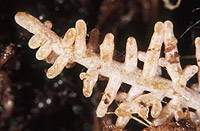
– Enlarged view – |
| • references | |
| Brand F (1989) Studies on ectomycorrhizae XXI. Beech ectomycorrhizae and rhizomorphs of Xerocomus chrysenteron (Boletales). Nova Hedwigia 48: 469-483. Brand F (1989) Xerocomus chrysenteron. In Agerer R (ed) Colour Atlas of Ectomycorrhizae, plate 34, Einhorn-Verlag, Schwäbisch Gmünd. Hofmann H-P (1989) The ultrastructure of natural beech ectomycorrhizae and rhizomorphs of the basidiomycete Xerocomus chrysenteron (Bull.: St. Amans) Quel. Nova Hedwigia 48: 455-468. |
|
| • length | |
| 0 mm | Lower value of unspecified range (could be µ-s.d., but not known) |
| 3.5 mm | Upper value of unspecified range (could be µ+s.d., but not known) |
| • ramification presence-type | |
| monopodial-pyramidal | |
| or | coralloid |
| • ramification orders | |
| 0 | Lower value of unspecified range (could be µ-s.d., but not known) |
| 2 | Upper value of unspecified range (could be µ+s.d., but not known) |
| • main axis diameter | |
| 0.5 mm | Lower value of unspecified range (could be µ-s.d., but not known) |
| 0.6 mm | Upper value of unspecified range (could be µ+s.d., but not known) |
| • rhizomorphs as stout, short, conical structures presence-abundance | |
| absent | |
| • rhizomorphs presence | |
| present | |
| • rhizomorphs frequency | |
| infrequent | |
| • exploration type | |
| long distance | |
| • shape | |
| straight | |
| or | bent |
| • shape {of distal end} | |
| not inflated, cylindric | |
| • length | |
| 0 mm | Lower value of unspecified range (could be µ-s.d., but not known) |
| 1.5 mm | Upper value of unspecified range (could be µ+s.d., but not known) |
| • diameter | |
| 0.35 mm | Lower value of unspecified range (could be µ-s.d., but not known) |
| 0.4 mm | Upper value of unspecified range (could be µ+s.d., but not known) |
| • colour | |
| brown | |
| or | ochre, yellowish brown |
| or | green |
| or | white |
| • very tip colour | |
| white | |
| • older parts colour | |
| brown | |
| or | ochre, yellowish brown |
| or | green |
| or | white |
| • mantle cortical cells visibility | |
| not visible | |
| • mantle {distinct} surface visibility | |
| present | |
| • mantle transparency | |
| not transparent | |
| • mantle laticifers visibility | |
| absent | |
| • mantle dots presence-colour | |
| absent | |
| • mantle carbonizing presence | |
| absent | |
| • mantle surface {in general} habit | |
| silvery | |
| or | smooth |
| • emanating hyphae presence | |
| absent | |
| or | present |
| • emanating hyphae abundance | |
| infrequent | |
| • diameter | |
| 0 mm | Lower value of unspecified range (could be µ-s.d., but not known) |
| 0.3 mm | Upper value of unspecified range (could be µ+s.d., but not known) |
| • cross-section shape | |
| round or roundish | |
| • colour | |
| concolourous to mantle | |
| or | yellowish |
| or | whitish |
| • ramification kind-frequency | |
| frequently, at restricted points | |
| • connection to mantle kind | |
| distinct | |
| • origin location | |
| proximal | |
| • margin habit | |
| smooth | |
| • dimorphism presence | |
| absent | |
| • presence | |
| absent | |
| • emanating elements presence-type | |
| rhizomorphs | |
| • presence | |
| absent | |
| • organisation | |
| plectenchymatous | |
| • mantle type | |
| ring-like arrangement of hyphal bundles (type A) | |
| • hyphae hyphal junctions angle {between} | |
| ca. 45° and less | |
| • septa thickness {relative to cell walls} | |
| as thick as walls | |
| • septa clamps presence | |
| absent | |
| • cell shape | |
| cylindric, constricted at septa | |
| • cell pigment location-colour | |
| plasmatically greenish | |
| and | plasmatically brownish |
| • cell diameter | |
| 3 µm | Mean (= average) |
| • cell wall thickness | |
| 0.2 µm | Mean (= average) |
| • cell wall surface habit | |
| smooth | |
| • drops of exuded pigment presence | |
| absent | |
| • organisation | |
| plectenchymatous | |
| • cell diameter | |
| 4 µm | Lower value of unspecified range (could be µ-s.d., but not known) |
| 6 µm | Upper value of unspecified range (could be µ+s.d., but not known) |
| 10 µm | Maximum value |
| • cell wall surface habit | |
| smooth | |
| • organisation | |
| plectenchymatous | |
| • hyphae arrangement | |
| without pattern | |
| or | ring-like |
| • septa clamps presence | |
| absent | |
| • cell diameter | |
| 3 µm | Lower value of unspecified range (could be µ-s.d., but not known) |
| 4 µm | Upper value of unspecified range (could be µ+s.d., but not known) |
| • mantle thickness {apart from tip} | |
| 40 µm | Lower value of unspecified range (could be µ-s.d., but not known) |
| 60 µm | Upper value of unspecified range (could be µ+s.d., but not known) |
| • mantle different layers presence | |
| discernable | |
| • outer mantle layer organisation | |
| plectenchymatous | |
| • outer mantle layer hyphae radially diameter | |
| 3 µm | Lower value of unspecified range (could be µ-s.d., but not known) |
| 8 µm | Upper value of unspecified range (could be µ+s.d., but not known) |
| • inner mantle layer organisation | |
| plectenchymatous | |
| • inner mantle layer hyphae radially diameter | |
| 3 µm | Lower value of unspecified range (could be µ-s.d., but not known) |
| 8 µm | Upper value of unspecified range (could be µ+s.d., but not known) |
| • presence | |
| absent | |
| • anatomy mantle longitudinal section cortical (epidermal) cells shape | |
| radially-oval to -elliptic, oriented obliquely | |
| • anatomy mantle longitudinal section cortical (epidermal) cells tangentially length | |
| 10 µm | Lower value of unspecified range (could be µ-s.d., but not known) |
| 20 µm | Upper value of unspecified range (could be µ+s.d., but not known) |
| • anatomy mantle longitudinal section cortical (epidermal) cells radially diameter | |
| 30 µm | Lower value of unspecified range (could be µ-s.d., but not known) |
| 55 µm | Upper value of unspecified range (could be µ+s.d., but not known) |
| • presence | |
| present | |
| • kind | |
| periepidermal | |
| • structure {in plan view} | |
| of palmetti type | |
| • mantle different layers presence | |
| discernible | |
| • outer mantle layer organisation | |
| plectenchymatous | |
| • outer mantle layer hyphae radially diameter | |
| 3 µm | Lower value of unspecified range (could be µ-s.d., but not known) |
| 8 µm | Upper value of unspecified range (could be µ+s.d., but not known) |
| • inner mantle layer organisation | |
| plectenchymatous | |
| • inner mantle layer hyphae radially diameter | |
| 3 µm | Lower value of unspecified range (could be µ-s.d., but not known) |
| 8 µm | Upper value of unspecified range (could be µ+s.d., but not known) |
| • presence | |
| absent | |
| • anatomy mantle cross-section cortical (epidermal) cells shape | |
| round | |
| or | radially-oval to -elliptic |
| • anatomy mantle cross-section cortical (epidermal) cells tangentially length | |
| 12 µm | Lower value of unspecified range (could be µ-s.d., but not known) |
| 25 µm | Upper value of unspecified range (could be µ+s.d., but not known) |
| • anatomy mantle cross-section cortical (epidermal) cells radially diameter | |
| 15 µm | Lower value of unspecified range (could be µ-s.d., but not known) |
| 25 µm | Upper value of unspecified range (could be µ+s.d., but not known) |
| • presence | |
| present | |
| • kind | |
| in tow rows, not reaching endodermis | |
| • anatomy mantle cross-section hyphal cells around cortical (epidermal) cells thickness | |
| 1.5 µm | Lower value of unspecified range (could be µ-s.d., but not known) |
| 2.5 µm | Upper value of unspecified range (could be µ+s.d., but not known) |
| • septal pores configuration | |
| dolipore-like structures | |
| • anastomoses type | |
| open, with a short bridge or bridge almost lacking | |
| • drops of exuded pigment presence | |
| absent | |
| • clamps presence | |
| absent | |
| • anatomy emanating elements emanating hyphae cell shape | |
| slightly constricted | |
| • anatomy emanating elements emanating hyphae cell shape {at distal end} | |
| simple | |
| • anatomy emanating elements emanating hyphae cell diameter | |
| 3 µm | Mean (= average) |
| • anatomy emanating elements emanating hyphae cell wall surface habit | |
| smooth | |
| or | without lens-shaped appositions |
| or | without spindle-shaped appositions |
| • anatomy emanating elements emanating hyphae cell wall thickness | |
| 0.2 µm | Mean (= average) |
| • anatomy emanating elements emanating hyphae cell wall thickness at tip {relative to remaining cell wall} | |
| thinner | |
| • anatomy emanating elements emanating hyphae cell wall {apart from tip} evenness | |
| even in thickness | |
| • type | |
| highly differentiated; thick hyphae forming mostly a core, septa often partially or completely dissolved (type F) |
|
| • nodia presence | |
| present | |
| • internal nodia presence | |
| present | |
| • gelatinous matrix presence | |
| absent | |
| • a "ball" of intertwined, ramified, thin hyphae presence | |
| absent | |
| • ampullate, trumpet-like inflated presence | |
| absent | |
| • anatomy emanating elements rhizomorphs hyphae ampullate, trumpet-like inflated presence | |
| absent | |
| • anatomy emanating elements rhizomorphs hyphae ampullate, trumpet-like inflated presence | |
| absent | |
| • anatomy emanating elements rhizomorphs hyphae ampullate, trumpet-like inflated presence | |
| absent | |
| • anatomy emanating elements rhizomorphs hyphae ampullate, trumpet-like inflated presence | |
| absent | |
| • presence | |
| absent | |
| • {of ectomycorrhiza former} presence | |
| absent | |
| • {of foreign origin} presence | |
| absent | |
| • number {per cell} | |
| 2 | Mean (= average) |
| • shape | |
| round | |
| • diameter | |
| 1 µm | Lower value of unspecified range (could be µ-s.d., but not known) |
| 1.8 µm | Upper value of unspecified range (could be µ+s.d., but not known) |
| • length | |
| 1 µm | Lower value of unspecified range (could be µ-s.d., but not known) |
| 1.8 µm | Upper value of unspecified range (could be µ+s.d., but not known) |
| • septal pores type | |
| as dolipores with perforated parenthesome | |
| • mantle matrix presence-kind | |
| transparent | |
| • whole mycorrhizae UV 254 nm colour-presence | |
| absent | |
| • whole mycorrhizae UV 366 nm colour-presence | |
| absent | |
| • mantle in section UV-filter 340-380 nm presence | |
| present | |
| • substrate | |
| in organic layer | |
| or | in organically enriched mineral horizon |
| • geographic occurrence continent | |
| Europe | |
| • knowledge about association with foreign fruitbodies presence | |
| unknown | |
| • plant family | |
| Fagaceae | |
| • plant genus | |
| Fagus | |
| • plant habitat kind | |
| forests, woods | |
| • family | |
| Boletaceae | |
| • subfamily | |
| Boletaceae subf. Xerocomoideae | |
| • fruitbodies growth habit | |
| epigeous | |
| or | pileate-porioid |
| • public notes | |
| Cell walls of middle mantle layer hyphae slightly thick; autofluorescence of rhizomorphs with UV-filter showing central hyphae bluish and surrounding hyphae pale ochre; distance of nuclei variable. | |