|
|
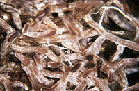
– Enlarged view – |
| • references | |
| Müller W, Agerer R (1990) Studien an Ektomykorrhizen XXIX. Drei Mykorrhizen aus der Leccinum-scabrum-Gruppe. Nova Hedwigia 51: 381-410. | |
| • length | |
| 0 mm | Lower value of unspecified range (could be µ-s.d., but not known) |
| 30 mm | Upper value of unspecified range (could be µ+s.d., but not known) |
| • ramification presence-type | |
| monopodial-pinnate | |
| or | monopodial-pyramidal |
| or | coralloid |
| or | braid-like |
| • ramification orders | |
| 0 | Lower value of unspecified range (could be µ-s.d., but not known) |
| 2 | Upper value of unspecified range (could be µ+s.d., but not known) |
| • main axis diameter | |
| 0.4 mm | Mean (= average) |
| • rhizomorphs as stout, short, conical structures presence-abundance | |
| absent | |
| • rhizomorphs as short mycorrhiza-like outgrowths with blunt tips presence | |
| absent | |
| • rhizomorphs presence | |
| present | |
| • rhizomorphs frequency | |
| abundant | |
| • exploration type | |
| long distance | |
| • shape | |
| bent | |
| or | tortuous |
| • shape {of distal end} | |
| not inflated, cylindric | |
| • length | |
| 0 mm | Lower value of unspecified range (could be µ-s.d., but not known) |
| 3 mm | Upper value of unspecified range (could be µ+s.d., but not known) |
| • diameter | |
| 0.3 mm | Mean (= average) |
| • colour | |
| dark brown | |
| or | brown |
| or | ochre, yellowish brown |
| or | red |
| or | blue |
| • mantle cortical cells visibility | |
| not visible | |
| • mantle {distinct} surface visibility | |
| present | |
| • mantle transparency | |
| not transparent | |
| • mantle laticifers visibility | |
| absent | |
| • mantle dots presence-colour | |
| absent | |
| • mantle carbonizing presence | |
| absent | |
| • mantle surface {in general} habit | |
| silvery | |
| or | not smooth |
| • mantle surface {in detail} kind | |
| densely cottony | |
| • emanating hyphae presence | |
| present | |
| • emanating hyphae abundance | |
| abundant | |
| • emanating hyphae distribution | |
| not specifically distributed | |
| • diameter | |
| 0 mm | Lower value of unspecified range (could be µ-s.d., but not known) |
| 0.3 mm | Upper value of unspecified range (could be µ+s.d., but not known) |
| • cross-section shape | |
| round or roundish | |
| • colour | |
| blue | |
| or | green |
| or | white |
| • ramification kind-frequency | |
| frequently, at restricted points | |
| • connection to mantle kind | |
| distinct | |
| • margin habit | |
| hairy | |
| • dimorphism presence | |
| absent | |
| • presence | |
| absent | |
| • emanating elements presence-type | |
| rhizomorphs | |
| or | cystidia |
| • emanating elements cystidia location | |
| on outer mantle layer | |
| • blue granules presence | |
| absent | |
| • presence | |
| absent | |
| • organisation | |
| plectenchymatous | |
| • mantle type | |
| ring-like arrangement of hyphal bundles (type A) | |
| • hyphal system kind | |
| undifferentiated | |
| • hyphae hyphal junctions angle {between} | |
| ca. 90° | |
| and | ca. 120° |
| • septa thickness {relative to cell walls} | |
| as thick as walls | |
| • septa clamps presence | |
| absent | |
| • cell shape | |
| cylindric, not constricted at septa | |
| • cell pigment location-colour | |
| absent | |
| • cell diameter | |
| 2.5 µm | Lower value of unspecified range (could be µ-s.d., but not known) |
| 5 µm | Upper value of unspecified range (could be µ+s.d., but not known) |
| • cell wall thickness | |
| 0.2 µm | Mean (= average) |
| • cell wall surface habit | |
| smooth | |
| and | rough |
| • drops of exuded pigment presence | |
| absent | |
| • organisation | |
| plectenchymatous | |
| • hyphae arrangement | |
| plectenchymatous, ring-like | |
| • cell pigment location-colour | |
| colourless | |
| • cell diameter | |
| 4 µm | Lower value of unspecified range (could be µ-s.d., but not known) |
| 5 µm | Upper value of unspecified range (could be µ+s.d., but not known) |
| • cell contents presence-kind | |
| absent | |
| • cell wall thickness | |
| 0.2 µm | Mean (= average) |
| • cell wall surface habit | |
| smooth | |
| • organisation | |
| plectenchymatous with pseudoparenchymatous nests of cells | |
| • hyphae arrangement | |
| ring-like | |
| • septa clamps presence | |
| absent | |
| • cell pigment location-colour | |
| absent | |
| • cell diameter | |
| 3 µm | Lower value of unspecified range (could be µ-s.d., but not known) |
| 5 µm | Upper value of unspecified range (could be µ+s.d., but not known) |
| • cell contents presence-kind | |
| absent | |
| • mantle thickness {apart from tip} | |
| 25 µm | Lower value of unspecified range (could be µ-s.d., but not known) |
| 35 µm | Upper value of unspecified range (could be µ+s.d., but not known) |
| • mantle different layers presence | |
| discernable | |
| • outer mantle layer organisation | |
| plectenchymatous | |
| • outer mantle layer hyphae radially diameter | |
| 4 µm | Lower value of unspecified range (could be µ-s.d., but not known) |
| 5 µm | Upper value of unspecified range (could be µ+s.d., but not known) |
| • inner mantle layer organisation | |
| plectenchymatous | |
| • inner mantle layer hyphae radially diameter | |
| 4 µm | Lower value of unspecified range (could be µ-s.d., but not known) |
| 5 µm | Upper value of unspecified range (could be µ+s.d., but not known) |
| • presence | |
| absent | |
| • anatomy mantle longitudinal section cortical (epidermal) cells shape | |
| tangentially-oval to -elliptic or -cylindrical, and oriented obliquely | |
| • anatomy mantle longitudinal section cortical (epidermal) cells tangentially length | |
| 12 µm | Lower value of unspecified range (could be µ-s.d., but not known) |
| 35 µm | Upper value of unspecified range (could be µ+s.d., but not known) |
| • anatomy mantle longitudinal section cortical (epidermal) cells radially diameter | |
| 50 µm | Lower value of unspecified range (could be µ-s.d., but not known) |
| 75 µm | Upper value of unspecified range (could be µ+s.d., but not known) |
| • presence | |
| present | |
| • kind | |
| paraepidermal | |
| • anatomy mantle longitudinal section hyphal cells around tannin cells thickness | |
| 2 µm | Lower value of unspecified range (could be µ-s.d., but not known) |
| 5 µm | Upper value of unspecified range (could be µ+s.d., but not known) |
| 13 µm | Maximum value |
| • anatomy mantle longitudinal section hyphal cells around cortical (epidermal) cells shape | |
| beaded | |
| • structure {in plan view} | |
| of palmetti type | |
| • mantle different layers presence | |
| discernible | |
| • outer mantle layer organisation | |
| plectenchymatous | |
| • outer mantle layer hyphae radially diameter | |
| 4 µm | Lower value of unspecified range (could be µ-s.d., but not known) |
| 5 µm | Upper value of unspecified range (could be µ+s.d., but not known) |
| • inner mantle layer organisation | |
| plectenchymatous | |
| • inner mantle layer hyphae radially diameter | |
| 4 µm | Lower value of unspecified range (could be µ-s.d., but not known) |
| 5 µm | Upper value of unspecified range (could be µ+s.d., but not known) |
| • presence | |
| absent | |
| • anatomy mantle cross-section cortical (epidermal) cells tangentially length | |
| 15 µm | Lower value of unspecified range (could be µ-s.d., but not known) |
| 40 µm | Upper value of unspecified range (could be µ+s.d., but not known) |
| • anatomy mantle cross-section cortical (epidermal) cells radially diameter | |
| 50 µm | Lower value of unspecified range (could be µ-s.d., but not known) |
| 70 µm | Upper value of unspecified range (could be µ+s.d., but not known) |
| • anatomy mantle cross-section hyphal cells around cortical (epidermal) cells shape | |
| beaded | |
| • anatomy mantle cross-section hyphal cells around cortical (epidermal) cells thickness | |
| 2 µm | Lower value of unspecified range (could be µ-s.d., but not known) |
| 5 µm | Upper value of unspecified range (could be µ+s.d., but not known) |
| 13 µm | Maximum value |
| • anatomy mantle cross-section hyphal rows around cortical (epidermal) cells number | |
| one | |
| • structure {in plan view} | |
| of palmetti type | |
| • intrahyphal hyphae presence | |
| absent | |
| • backwards-oriented clamps presence | |
| absent | |
| • anastomoses type | |
| open, with a short bridge or bridge almost lacking | |
| • type | |
| as acanthocystidia (type P) | |
| • shape | |
| not striking | |
| • cell pigment location-colour | |
| absent | |
| • drops of exuded pigment presence | |
| absent | |
| • presence | |
| present | |
| • kind | |
| approximately 90° | |
| or | Y-shaped |
| • side-branches at septum number | |
| one side-branch at septum | |
| or | two true (at one height) side-branches at septum |
| • clamps presence | |
| absent | |
| • clamps hole presence | |
| absent | |
| • clamps blister-like structure {at basis} presence | |
| absent | |
| • anatomy emanating elements emanating hyphae cell shape | |
| even | |
| • anatomy emanating elements emanating hyphae cell wall surface habit | |
| smooth | |
| or | rough of warts |
| or | without lens-shaped appositions |
| or | without spindle-shaped appositions |
| • anatomy emanating elements emanating hyphae cell wall surface structures shape | |
| hemispherical warts | |
| • type | |
| highly differentiated; thick hyphae forming mostly a core, septa often partially or completely dissolved (type F) |
|
| • nodia presence | |
| present | |
| • internal nodia presence | |
| absent | |
| • gelatinous matrix presence | |
| absent | |
| • gelatinized hyphae presence | |
| absent | |
| • cup-like structures on surface presence | |
| absent | |
| • conical young side-branches presence | |
| present | |
| • a "ball" of intertwined, ramified, thin hyphae presence | |
| absent | |
| • ampullate, trumpet-like inflated presence | |
| absent | |
| • anatomy emanating elements rhizomorphs hyphae ampullate, trumpet-like inflated presence | |
| absent | |
| • anatomy emanating elements rhizomorphs hyphae ampullate, trumpet-like inflated presence | |
| absent | |
| • anatomy emanating elements rhizomorphs hyphae ampullate, trumpet-like inflated presence | |
| absent | |
| • anatomy emanating elements rhizomorphs hyphae ampullate, trumpet-like inflated presence | |
| absent | |
| • presence | |
| absent | |
| • {of ectomycorrhiza former} presence | |
| present | |
| • {of ectomycorrhiza former} abundance | |
| occasionally present | |
| • shape | |
| as normal hyphae | |
| or | globular |
| • number {per cell} | |
| 2 | Mean (= average) |
| • shape | |
| round | |
| • diameter | |
| 1.5 µm | Mean (= average) |
| • length | |
| 1.5 µm | Mean (= average) |
| • substrate | |
| below mosses | |
| • geographic occurrence continent | |
| Europe | |
| • knowledge about association with foreign fruitbodies presence | |
| unknown | |
| • plant family | |
| Betulaceae | |
| • plant genus | |
| Betula | |
| • plant habitat kind | |
| forests, woods | |
| • family | |
| Boletaceae | |
| • subfamily | |
| Boletaceae subf. Boletoideae | |
| • fruitbodies growth habit | |
| epigeous | |
| or | pileate-porioid |
| • public notes | |
| Mycorrhizal systems densely enveloped by a web of hyphae; mycorrhizal ends often hook-like, ochre, red-brown to dark brown, in water darker with a bluish tint, very tips can be darker than remaining parts; rhizomorphs dark green-blue where injured; mantle surface with acanthohyphae; Hartig net in section with inflated, beaded cells up to 13 um in diam.; mantle in sulfo-vanillin slightly reddish, in FeSO4 slightly darker, in brillant-cresyl-blue blue, in acid fuchsin red. | |