|
|
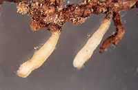
– Enlarged view – |
| • references | |
| Berg B (1989) Charakterisierung und Vergleich von Ektomykorrhizen gekalkter Fichtenbestände. Diss Univ München. Berg B, Gronboach E (1989) Piceirhiza gelatinosa. In Agerer R (ed) Colour Atlas of Ectomycorrhizae, plate 30, Einhorn-Verlag, Schwäbisch Gmünd. Franz F (1994) Ektomykorrhizen der Fichte: Identifizierung, Ultrastruktur und Mikroelementanalyse (EELS, ESI). Diss Univ Bayreuth. Gronbach E, Agerer R (1986) Charakterisierung und Inventur der Fichten-Mykorrhizen im Höglwald und deren Reaktion auf saure Beregnung. Forstwiss Cbl 105: 329-335. Haug I (1987) Licht- und elektronenmikroskopische Untersuchungen an Mykorrhizen von Fichtenbeständen im Schwarzwald. Diss Univ Tübingen. Haug I (1989) Intercellular infection in the meristematic region of Piceirhiza gelatinosa mycorrhizas. New Phytol 111: 203-207. Haug I, Oberwinkler F (1987) Some distinctive types of spruce mycorrhizae. Trees 1: 172-188. Haug I, Pritsch K (1992) Ectomycorrhizal types of spruce (Picea abies (L.) Karst.) in the Black Forest. A microscopical atlas. Kernforschungszentrum Karlsruhe. |
|
| • length | |
| 0 mm | Lower value of unspecified range (could be µ-s.d., but not known) |
| 6 mm | Upper value of unspecified range (could be µ+s.d., but not known) |
| • ramification presence-type | |
| absent | |
| • ramification orders | |
| 0 | Mean (= average) |
| • abundance | |
| solitary or in small numbers | |
| • rhizomorphs as stout, short, conical structures presence-abundance | |
| absent | |
| • rhizomorphs as short mycorrhiza-like outgrowths with blunt tips presence | |
| absent | |
| • rhizomorphs presence | |
| absent | |
| • exploration type | |
| contact | |
| • shape | |
| straight | |
| or | bent |
| • shape {of distal end} | |
| not inflated, cylindric | |
| or | tapering |
| • length | |
| 0 mm | Lower value of unspecified range (could be µ-s.d., but not known) |
| 4 mm | Upper value of unspecified range (could be µ+s.d., but not known) |
| • diameter | |
| 0.2 mm | Minimum value |
| 0.45 mm | Lower value of unspecified range (could be µ-s.d., but not known) |
| 0.8 mm | Upper value of unspecified range (could be µ+s.d., but not known) |
| 0.9 mm | Maximum value |
| • colour | |
| ochre, yellowish brown | |
| or | yellow |
| or | white |
| • very tip colour | |
| dark brown | |
| or | white |
| or | whitish |
| • older parts colour | |
| ochre, yellowish brown | |
| or | yellow |
| • mantle {distinct} surface visibility | |
| present | |
| • mantle laticifers visibility | |
| absent | |
| • mantle dots presence-colour | |
| absent | |
| • mantle carbonizing presence | |
| absent | |
| • mantle surface {in general} habit | |
| shiny | |
| or | smooth |
| • emanating hyphae presence | |
| present | |
| • emanating hyphae abundance | |
| infrequent | |
| • presence | |
| absent | |
| • presence | |
| absent | |
| • matrix presence | |
| present | |
| • matrix location | |
| outer mantle layer {apart from tip} | |
| or | outer mantle layer {of ectomycorrhizal tip} |
| or | middle mantle layer |
| or | inner mantle layer |
| • organisation | |
| plectenchymatous | |
| • mantle type | |
| hyphae rather irregularly arranged and no special pattern discernible (type B) | |
| and | gelatinous matrix between the hyphae (type C) |
| • matrix kind | |
| gelatinous | |
| • hyphal system kind | |
| meandering | |
| • cell shape | |
| cylindric, constricted at septa | |
| and | ampullate at both sides of septum |
| • cell pigment location-colour | |
| absent | |
| • cell diameter | |
| 1 µm | Minimum value |
| 2 µm | Lower value of unspecified range (could be µ-s.d., but not known) |
| 3.5 µm | Upper value of unspecified range (could be µ+s.d., but not known) |
| 5 µm | Maximum value |
| • cell length | |
| 10 µm | Lower value of unspecified range (could be µ-s.d., but not known) |
| 60 µm | Upper value of unspecified range (could be µ+s.d., but not known) |
| • cell wall thickness | |
| 0.2 µm | Mean (= average) |
| • cell wall surface habit | |
| smooth | |
| • drops of exuded pigment presence | |
| absent | |
| • organisation | |
| plectenchymatous | |
| • matrix kind | |
| gelatinous | |
| • hyphae arrangement | |
| plectenchymatous, ring-like | |
| • cell pigment location-colour | |
| colourless | |
| • cell diameter | |
| 2 µm | Lower value of unspecified range (could be µ-s.d., but not known) |
| 3 µm | Upper value of unspecified range (could be µ+s.d., but not known) |
| 4 µm | Maximum value |
| • cell length | |
| 10 µm | Lower value of unspecified range (could be µ-s.d., but not known) |
| 60 µm | Upper value of unspecified range (could be µ+s.d., but not known) |
| • cell wall thickness | |
| 0.2 µm | Mean (= average) |
| • cell wall surface habit | |
| smooth | |
| • organisation | |
| plectenchymatous | |
| • matrix kind | |
| gelatinous | |
| • hyphae arrangement | |
| ring-like | |
| • septa clamps presence | |
| absent | |
| • cell pigment location-colour | |
| absent | |
| • cell diameter | |
| 2 µm | Lower value of unspecified range (could be µ-s.d., but not known) |
| 3 µm | Upper value of unspecified range (could be µ+s.d., but not known) |
| 5 µm | Maximum value |
| • anatomy mantle outer mantle layer {of ectomycorrhizal tip} organisation | |
| like other parts of mantle | |
| • anatomy mantle outer mantle layer {of ectomycorrhizal tip} matrix kind | |
| gelatinous | |
| • anatomy mantle outer mantle layer {of ectomycorrhizal tip} hyphae diameter | |
| 2 µm | Lower value of unspecified range (could be µ-s.d., but not known) |
| 3 µm | Upper value of unspecified range (could be µ+s.d., but not known) |
| 4 µm | Maximum value |
| • mantle thickness {apart from tip} | |
| 23 µm | Minimum value |
| 32 µm | Lower value of unspecified range (could be µ-s.d., but not known) |
| 55 µm | Upper value of unspecified range (could be µ+s.d., but not known) |
| 72 µm | Maximum value |
| • mantle different layers presence | |
| not discernable | |
| • outer mantle layer organisation | |
| plectenchymatous | |
| • middle mantle layer organisation | |
| plectenchymatous | |
| • inner mantle layer organisation | |
| plectenchymatous | |
| • unlayered mantle hyphae tangentially length | |
| 1 µm | Minimum value |
| 2 µm | Lower value of unspecified range (could be µ-s.d., but not known) |
| 4 µm | Upper value of unspecified range (could be µ+s.d., but not known) |
| 5 µm | Maximum value |
| • unlayered mantle hyphae radially diameter | |
| 1 µm | Minimum value |
| 2 µm | Lower value of unspecified range (could be µ-s.d., but not known) |
| 3 µm | Upper value of unspecified range (could be µ+s.d., but not known) |
| 4 µm | Maximum value |
| • presence | |
| present | |
| • rows number | |
| 1 | Lower value of unspecified range (could be µ-s.d., but not known) |
| 3 | Upper value of unspecified range (could be µ+s.d., but not known) |
| • shape | |
| tangentially-oval, -elliptic or -cylindrical, and oriented in parallel to root axis | |
| • tangentially length | |
| 20 µm | Lower value of unspecified range (could be µ-s.d., but not known) |
| 70 µm | Upper value of unspecified range (could be µ+s.d., but not known) |
| 100 µm | Maximum value |
| • radially diameter | |
| 2 µm | Lower value of unspecified range (could be µ-s.d., but not known) |
| 24 µm | Upper value of unspecified range (could be µ+s.d., but not known) |
| 26 µm | Maximum value |
| • mean tangenial length TCt | |
| 53.6 µm | Mean (= average) |
| • mean shape-ratio TCq | |
| 4.5 | Mean (= average) |
| • anatomy mantle longitudinal section cortical (epidermal) cells shape | |
| round | |
| or | tangentially-oval to -elliptic or -cylindrical, and oriented in parallel to root axis |
| • anatomy mantle longitudinal section cortical (epidermal) cells tangentially length | |
| 10 µm | Minimum value |
| 25 µm | Lower value of unspecified range (could be µ-s.d., but not known) |
| 50 µm | Upper value of unspecified range (could be µ+s.d., but not known) |
| 60 µm | Maximum value |
| • anatomy mantle longitudinal section cortical (epidermal) cells radially diameter | |
| 10 µm | Lower value of unspecified range (could be µ-s.d., but not known) |
| 36 µm | Upper value of unspecified range (could be µ+s.d., but not known) |
| 40 µm | Maximum value |
| • anatomy mantle longitudinal section cortical (epidermal) cells mean tangential length CCt (ECt) | |
| 37.9 µm | Mean (= average) |
| • anatomy mantle longitudinal section cortical (epidermal) cells mean shape-ratio CCq (ECq) | |
| 1.4 | Mean (= average) |
| • presence | |
| present | |
| • kind | |
| protruding towards endodermis | |
| • anatomy mantle longitudinal section hyphal cells around tannin cells shape | |
| roundish | |
| • anatomy mantle longitudinal section hyphal cells around tannin cells thickness | |
| 2 µm | Lower value of unspecified range (could be µ-s.d., but not known) |
| 3 µm | Upper value of unspecified range (could be µ+s.d., but not known) |
| 4 µm | Maximum value |
| • anatomy mantle longitudinal section hyphal rows around tannin cells number | |
| one | |
| • anatomy mantle longitudinal section hyphal cells around cortical (epidermal) cells shape | |
| roundish | |
| • anatomy mantle longitudinal section hyphal cells around cortical cells (epidermal) thickness | |
| 3 µm | Minimum value |
| 5 µm | Lower value of unspecified range (could be µ-s.d., but not known) |
| 15 µm | Upper value of unspecified range (could be µ+s.d., but not known) |
| 30 µm | Maximum value |
| • anatomy mantle longitudinal section hyphal rows around cortical (epidermal) cells number | |
| two | |
| or | several |
| • structure {in plan view} | |
| of palmetti type | |
| • lobes width | |
| 2 µm | Lower value of unspecified range (could be µ-s.d., but not known) |
| 3 µm | Upper value of unspecified range (could be µ+s.d., but not known) |
| • mantle different layers presence | |
| not discernible | |
| • outer mantle layer organisation | |
| plectenchymatous | |
| • middle mantle layer organisation | |
| plectenchymatous | |
| • inner mantle layer organisation | |
| plectenchymatous | |
| • unlayered mantle hyphae radially diameter | |
| 2 µm | Lower value of unspecified range (could be µ-s.d., but not known) |
| 3 µm | Upper value of unspecified range (could be µ+s.d., but not known) |
| 4 µm | Maximum value |
| • presence | |
| present | |
| • rows number | |
| 1 | Lower value of unspecified range (could be µ-s.d., but not known) |
| 2 | Upper value of unspecified range (could be µ+s.d., but not known) |
| 3 | Maximum value |
| • shape | |
| tangentially-oval to tangentially-elliptic | |
| • tangentially length | |
| 20 µm | Minimum value |
| 30 µm | Lower value of unspecified range (could be µ-s.d., but not known) |
| 72 µm | Upper value of unspecified range (could be µ+s.d., but not known) |
| 90 µm | Maximum value |
| • radially diameter | |
| 2 µm | Lower value of unspecified range (could be µ-s.d., but not known) |
| 14 µm | Upper value of unspecified range (could be µ+s.d., but not known) |
| 26 µm | Maximum value |
| • mean tangential length TCt | |
| 57 µm | Mean (= average) |
| • mean shape-ratio TCq | |
| 4.8 | Mean (= average) |
| • anatomy mantle cross-section cortical (epidermal) cells shape | |
| round | |
| or | tangentially-oval to tangentially-elliptic |
| • anatomy mantle cross-section cortical (epidermal) cells tangentially length | |
| 20 µm | Lower value of unspecified range (could be µ-s.d., but not known) |
| 60 µm | Upper value of unspecified range (could be µ+s.d., but not known) |
| 65 µm | Maximum value |
| • anatomy mantle cross-section cortical (epidermal) cells radially diameter | |
| 14 µm | Lower value of unspecified range (could be µ-s.d., but not known) |
| 36 µm | Upper value of unspecified range (could be µ+s.d., but not known) |
| 40 µm | Maximum value |
| • anatomy mantle cross-section cortical (epidermal) cells mean tangential length CCt | |
| 47.5 µm | Mean (= average) |
| • anatomy mantle cross-section cortical (epidermal) cells mean shape-ratio CCq | |
| 1.8 | Mean (= average) |
| • presence | |
| present | |
| • kind | |
| protruding towards endodermis | |
| • anatomy mantle cross-section hyphal cells around tannin cells shape | |
| roundish | |
| or | cylindrical |
| • anatomy mantle cross-section hyphal cells around tannin cells thickness | |
| 1 µm | Minimum value |
| 2 µm | Lower value of unspecified range (could be µ-s.d., but not known) |
| 4 µm | Upper value of unspecified range (could be µ+s.d., but not known) |
| 5 µm | Maximum value |
| • anatomy mantle cross-section hyphal rows around tannin cells number | |
| one | |
| or | two |
| or | three |
| • anatomy mantle cross-section hyphal cells around cortical (epidermal) cells shape | |
| roundish | |
| or | cylindrical |
| • anatomy mantle cross-section hyphal cells around cortical (epidermal) cells thickness | |
| 2 µm | Lower value of unspecified range (could be µ-s.d., but not known) |
| 15 µm | Upper value of unspecified range (could be µ+s.d., but not known) |
| 30 µm | Maximum value |
| • anatomy mantle cross-section hyphal rows around cortical (epidermal) cells number | |
| one | |
| or | two |
| or | several |
| • lobes width | |
| 2 µm | Lower value of unspecified range (could be µ-s.d., but not known) |
| 3 µm | Upper value of unspecified range (could be µ+s.d., but not known) |
| • structure {in plan view} | |
| of palmetti type | |
| • shape | |
| wavy | |
| or | not striking |
| • cell pigment location-colour | |
| absent | |
| • drops of exuded pigment presence | |
| absent | |
| • clamps presence | |
| present | |
| • clamps outline {in lateral view} | |
| less than a semicircle | |
| or | not constricted at contact point to subtending hyphal cell |
| • clamps width {relative to hypha in lateral view} | |
| thinner than | |
| • clamps blister-like structure {at basis} presence | |
| absent | |
| • presence | |
| present | |
| • abundance | |
| infrequent | |
| • distribution | |
| not specified | |
| • anatomy emanating elements emanating hyphae cell shape {at distal end} | |
| simple | |
| • anatomy emanating elements emanating hyphae cell diameter | |
| 1 µm | Minimum value |
| 2 µm | Lower value of unspecified range (could be µ-s.d., but not known) |
| 3 µm | Upper value of unspecified range (could be µ+s.d., but not known) |
| 5 µm | Maximum value |
| • anatomy emanating elements emanating hyphae cell length | |
| 10 µm | Lower value of unspecified range (could be µ-s.d., but not known) |
| 20 µm | Upper value of unspecified range (could be µ+s.d., but not known) |
| • anatomy emanating elements emanating hyphae cell wall surface habit | |
| smooth | |
| or | without lens-shaped appositions |
| or | without spindle-shaped appositions |
| • type | |
| lacking, only emanating hyphae present (type G) |
|
| • presence | |
| absent | |
| • {of ectomycorrhiza former} presence | |
| absent | |
| • number {per cell} | |
| 2 | Mean (= average) |
| • shape | |
| round | |
| or | oval |
| • diameter | |
| 2 µm | Lower value of unspecified range (could be µ-s.d., but not known) |
| 3 µm | Upper value of unspecified range (could be µ+s.d., but not known) |
| • length | |
| 2 µm | Lower value of unspecified range (could be µ-s.d., but not known) |
| 3 µm | Upper value of unspecified range (could be µ+s.d., but not known) |
| • septal pores type | |
| as dolipores with perforated parenthesome | |
| • mantle matrix presence-kind | |
| transparent | |
| • geographic occurrence continent | |
| Europe | |
| • plant family | |
| Pinaceae | |
| • plant genus | |
| Picea | |
| • plant habitat kind | |
| forests, woods | |
| • public notes | |
| Mycorrhizal ends rarely slightly bent, slightly yellowish, light yellow, sometimes amost white, older parts yellowish to ochre; inner mantle layers with labyrinthic pattern; in longitudinal section cortical cells roundish to tangentially-oval; lobes of Hartig net in plan view septate; cortical cells in cross-section roundish to tangentially-oval; in meristematic regions foreign intercellular hyphae may occur; autofluorescence of mantle with UV-filter whitish blue, with blue-filter yellowish green, with green-filter red; mantle with brillant-cresyl-blue blue and cytoplasm light violet, with toluidine blue cytoplasm violet blue and matrix violet blue, with anilin cell walls slightly greyish, with acid fuchsin cytoplasm light rosy, with ruthenium red cytoplasm light rosy and cell walls slightly greyish; nuclei two and more, close together but also distant. | |