|
|
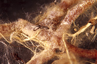
– Enlarged view – |
| • references | |
| Agerer R (1987-1995) Colour Atlas of Ectomycorrhizae. 1st-9th delivery. Einhorn, Schwäbisch Gmünd. Berg B (1989) Charakterisierung und Vergleich von Ektomykorrhizen gekalkter Fichtenbestände. Diss Univ München. Danielson RM, Pruden M (1989) The mycorrhizal status of urban spruce. Mycologia 81(3): 335-341. Haug I, Pritsch K (1992) Ectomycorrhizal types of spruce (Picea abies (L.) Karst.) in the Black Forest, a microscopical atlas. Kernforschungszentrum, Karlsruhe. Ingleby K, Mason PA, Last FT, Fleming LV (1990) Identification of ectomycorrhizas. ITE Research Publication no. 5. HMSO, London. Weiss M (1988) Ektomykorrhizen von Picea abies. Synthese, Ontogenie und Reaktion auf Umweltschadstoffe. Diss Univ München. Weiss M (1989) Amphinema byssoides. In Agerer R (ed) Colour Atlas of Ectomycorrhizae, plate 23, Einhorn-Verlag, Schwäbisch Gmünd. Weiss M (1990) Studien an Ektomykorrhizen XXV. Untersuchungen zur Ontogenie von Ektomykorrhizen an Picea abies. Nova Hedwigia 50: 361-393. Weiss M, Agerer R (1988) Studien an Ektomykorrhizen XII. Drei nichtidentifizierte Mykorrhizen an Picea abies (L.) Karst. aus einer Baumschule. Eur. J. For. Path. 18: 26-43. |
|
| • length | |
| 4 mm | Lower value of unspecified range (could be µ-s.d., but not known) |
| 10 mm | Upper value of unspecified range (could be µ+s.d., but not known) |
| • ramification presence-type | |
| monopodial-pinnate | |
| • ramification orders | |
| 0 | Lower value of unspecified range (could be µ-s.d., but not known) |
| 1 | Upper value of unspecified range (could be µ+s.d., but not known) |
| • abundance | |
| solitary or in small numbers | |
| • main axis diameter | |
| 0.4 mm | Lower value of unspecified range (could be µ-s.d., but not known) |
| 0.5 mm | Upper value of unspecified range (could be µ+s.d., but not known) |
| • rhizomorphs as stout, short, conical structures presence-abundance | |
| absent | |
| • rhizomorphs as short mycorrhiza-like outgrowths with blunt tips presence | |
| absent | |
| • rhizomorphs presence | |
| present | |
| • rhizomorphs frequency | |
| abundant | |
| • exploration type | |
| medium distance fringe | |
| • shape | |
| straight | |
| or | bent |
| • shape {of distal end} | |
| not inflated, cylindric | |
| • length | |
| 0 mm | Lower value of unspecified range (could be µ-s.d., but not known) |
| 3 mm | Upper value of unspecified range (could be µ+s.d., but not known) |
| 4 mm | Maximum value |
| • diameter | |
| 0.2 mm | Minimum value |
| 0.25 mm | Lower value of unspecified range (could be µ-s.d., but not known) |
| 0.4 mm | Upper value of unspecified range (could be µ+s.d., but not known) |
| 0.5 mm | Maximum value |
| • colour | |
| yellow | |
| • very tip colour | |
| whitish | |
| • older parts colour | |
| brown | |
| or | yellow |
| • mantle cortical cells visibility | |
| not visible | |
| • mantle {distinct} surface visibility | |
| absent | |
| • mantle transparency | |
| not transparent | |
| • mantle laticifers visibility | |
| absent | |
| • mantle dots presence-colour | |
| absent | |
| • mantle carbonizing presence | |
| absent | |
| • mantle surface {in general} habit | |
| silvery | |
| • mantle surface {in detail} kind | |
| densely woolly | |
| or | densely cottony |
| or | densely stringy |
| or | loosely stringy |
| • emanating hyphae presence | |
| present | |
| • emanating hyphae abundance | |
| abundant | |
| • emanating hyphae distribution | |
| not specifically distributed | |
| • diameter | |
| 0.008 mm | Lower value of unspecified range (could be µ-s.d., but not known) |
| 0.2 mm | Upper value of unspecified range (could be µ+s.d., but not known) |
| • cross-section shape | |
| round or roundish | |
| • colour | |
| yellow | |
| • ramification kind-frequency | |
| repeatedly into smaller filaments | |
| • connection to mantle kind | |
| fan-like | |
| • origin location | |
| not specific | |
| or | proximal |
| • margin habit | |
| densely enveloped by hyphae | |
| • dimorphism presence | |
| absent | |
| • presence | |
| absent | |
| • emanating elements presence-type | |
| rhizomorphs | |
| • blue granules presence | |
| absent | |
| • presence | |
| absent | |
| • organisation | |
| plectenchymatous | |
| • mantle type | |
| hyphae rather irregularly arranged and no special pattern discernible (type B) | |
| • hyphal system kind | |
| undifferentiated | |
| • septa thickness {relative to cell walls} | |
| as thick as walls | |
| • cell shape | |
| cylindric, not constricted at septa | |
| • cell contents presence-kind | |
| absent | |
| • cell diameter | |
| 2 µm | Lower value of unspecified range (could be µ-s.d., but not known) |
| 3 µm | Upper value of unspecified range (could be µ+s.d., but not known) |
| 4 µm | Maximum value |
| • cell length | |
| 20 µm | Lower value of unspecified range (could be µ-s.d., but not known) |
| 60 µm | Upper value of unspecified range (could be µ+s.d., but not known) |
| • cell wall thickness | |
| 0.2 µm | Mean (= average) |
| • cell wall surface habit | |
| rough | |
| and | with few crystals |
| • drops of exuded pigment presence | |
| absent | |
| • organisation | |
| plectenchymatous | |
| • hyphae arrangement | |
| plectenchymatous, without pattern | |
| • organisation | |
| plectenchymatous | |
| • hyphae arrangement | |
| without pattern | |
| • septa clamps presence | |
| absent | |
| • cell pigment location-colour | |
| absent | |
| • cell diameter | |
| 1 µm | Lower value of unspecified range (could be µ-s.d., but not known) |
| 4 µm | Upper value of unspecified range (could be µ+s.d., but not known) |
| • cell length | |
| 5 µm | Lower value of unspecified range (could be µ-s.d., but not known) |
| 60 µm | Upper value of unspecified range (could be µ+s.d., but not known) |
| • mantle thickness {apart from tip} | |
| 4 µm | Minimum value |
| 10 µm | Lower value of unspecified range (could be µ-s.d., but not known) |
| 24 µm | Upper value of unspecified range (could be µ+s.d., but not known) |
| 30 µm | Maximum value |
| • mantle different layers presence | |
| not discernable | |
| • outer mantle layer organisation | |
| plectenchymatous | |
| • middle mantle layer organisation | |
| plectenchymatous | |
| • inner mantle layer organisation | |
| plectenchymatous | |
| • unlayered mantle hyphae radially diameter | |
| 1.5 µm | Lower value of unspecified range (could be µ-s.d., but not known) |
| 3 µm | Upper value of unspecified range (could be µ+s.d., but not known) |
| 4 µm | Maximum value |
| • presence | |
| present | |
| • rows number | |
| 2 | Lower value of unspecified range (could be µ-s.d., but not known) |
| 3 | Upper value of unspecified range (could be µ+s.d., but not known) |
| 4 | Maximum value |
| • shape | |
| tangentially-oval, -elliptic or -cylindrical, and oriented in parallel to root axis | |
| • tangentially length | |
| 40 µm | Minimum value |
| 60 µm | Lower value of unspecified range (could be µ-s.d., but not known) |
| 120 µm | Upper value of unspecified range (could be µ+s.d., but not known) |
| • radially diameter | |
| 4 µm | Minimum value |
| 6 µm | Lower value of unspecified range (could be µ-s.d., but not known) |
| 10 µm | Upper value of unspecified range (could be µ+s.d., but not known) |
| 15 µm | Maximum value |
| • mean tangenial length TCt | |
| 78.8 µm | Mean (= average) |
| • mean shape-ratio TCq | |
| 9.3 | Mean (= average) |
| • anatomy mantle longitudinal section cortical (epidermal) cells shape | |
| tangentially-oval to -elliptic or -cylindrical, and oriented in parallel to root axis | |
| • anatomy mantle longitudinal section cortical (epidermal) cells tangentially length | |
| 35 µm | Minimum value |
| 45 µm | Lower value of unspecified range (could be µ-s.d., but not known) |
| 100 µm | Upper value of unspecified range (could be µ+s.d., but not known) |
| • anatomy mantle longitudinal section cortical (epidermal) cells radially diameter | |
| 5 µm | Minimum value |
| 10 µm | Lower value of unspecified range (could be µ-s.d., but not known) |
| 30 µm | Upper value of unspecified range (could be µ+s.d., but not known) |
| 40 µm | Maximum value |
| • anatomy mantle longitudinal section cortical (epidermal) cells mean tangential length CCt (ECt) | |
| 62.7 µm | Mean (= average) |
| • anatomy mantle longitudinal section cortical (epidermal) cells mean shape-ratio CCq (ECq) | |
| 4.9 | Mean (= average) |
| • presence | |
| present | |
| • kind | |
| protruding towards endodermis | |
| • anatomy mantle longitudinal section hyphal cells around tannin cells shape | |
| roundish | |
| • anatomy mantle longitudinal section hyphal rows around tannin cells number | |
| one | |
| • anatomy mantle longitudinal section hyphal cells around cortical (epidermal) cells shape | |
| roundish | |
| • anatomy mantle longitudinal section hyphal rows around cortical (epidermal) cells number | |
| one | |
| • structure {in plan view} | |
| of palmetti type | |
| • lobes width | |
| 1 µm | Lower value of unspecified range (could be µ-s.d., but not known) |
| 4 µm | Upper value of unspecified range (could be µ+s.d., but not known) |
| • mantle different layers presence | |
| not discernible | |
| • outer mantle layer organisation | |
| plectenchymatous | |
| • middle mantle layer organisation | |
| plectenchymatous | |
| • inner mantle layer organisation | |
| plectenchymatous | |
| • unlayered mantle hyphae radially diameter | |
| 1.5 µm | Lower value of unspecified range (could be µ-s.d., but not known) |
| 3 µm | Upper value of unspecified range (could be µ+s.d., but not known) |
| 4 µm | Maximum value |
| • presence | |
| present | |
| • rows number | |
| 2 | Lower value of unspecified range (could be µ-s.d., but not known) |
| 3 | Upper value of unspecified range (could be µ+s.d., but not known) |
| 4 | Maximum value |
| • shape | |
| tangentially-oval to tangentially-elliptic | |
| • tangentially length | |
| 15 µm | Lower value of unspecified range (could be µ-s.d., but not known) |
| 45 µm | Upper value of unspecified range (could be µ+s.d., but not known) |
| 60 µm | Maximum value |
| • radially diameter | |
| 4 µm | Minimum value |
| 6 µm | Lower value of unspecified range (could be µ-s.d., but not known) |
| 10 µm | Upper value of unspecified range (could be µ+s.d., but not known) |
| 15 µm | Maximum value |
| • mean tangential length TCt | |
| 39.3 µm | Mean (= average) |
| • mean shape-ratio TCq | |
| 4.6 | Mean (= average) |
| • anatomy mantle cross-section cortical (epidermal) cells shape | |
| round | |
| or | tangentially-oval to tangentially-elliptic |
| • anatomy mantle cross-section cortical (epidermal) cells tangentially length | |
| 18 µm | Lower value of unspecified range (could be µ-s.d., but not known) |
| 30 µm | Upper value of unspecified range (could be µ+s.d., but not known) |
| 40 µm | Maximum value |
| • anatomy mantle cross-section cortical (epidermal) cells radially diameter | |
| 5 µm | Minimum value |
| 10 µm | Lower value of unspecified range (could be µ-s.d., but not known) |
| 20 µm | Upper value of unspecified range (could be µ+s.d., but not known) |
| 25 µm | Maximum value |
| • anatomy mantle cross-section cortical (epidermal) cells mean tangential length CCt | |
| 23.9 µm | Mean (= average) |
| • anatomy mantle cross-section cortical (epidermal) cells mean shape-ratio CCq | |
| 1.9 | Mean (= average) |
| • anatomy mantle cross-section hyphal cells around tannin cells shape | |
| roundish | |
| • anatomy mantle cross-section hyphal cells around tannin cells thickness | |
| 1 µm | Lower value of unspecified range (could be µ-s.d., but not known) |
| 5 µm | Upper value of unspecified range (could be µ+s.d., but not known) |
| • anatomy mantle cross-section hyphal rows around tannin cells number | |
| one | |
| • anatomy mantle cross-section hyphal cells around cortical (epidermal) cells shape | |
| roundish | |
| • anatomy mantle cross-section hyphal cells around cortical (epidermal) cells thickness | |
| 2 µm | Lower value of unspecified range (could be µ-s.d., but not known) |
| 5 µm | Upper value of unspecified range (could be µ+s.d., but not known) |
| • anatomy mantle cross-section hyphal rows around cortical (epidermal) cells number | |
| one | |
| • intrahyphal hyphae presence | |
| absent | |
| • anastomoses type | |
| closed by a clamp, with a short bridge or bridge almost lacking (contact-clamp) | |
| • anastomoses surface habit | |
| rough or with crystals | |
| • anastomoses anastomosal bridge thickness {relative to hyphae} | |
| as thick as | |
| • anastomoses location | |
| not specified | |
| • shape | |
| wavy | |
| • cell pigment location-colour | |
| absent | |
| or | membranaceously yellowish |
| • drops of exuded pigment presence | |
| absent | |
| • presence | |
| present | |
| • kind | |
| approximately 90° | |
| or | Y-shaped |
| • distance {from septum} | |
| one or two hyphal diam. below the septum | |
| or | in considerable distance from the septum |
| • clamps presence | |
| present | |
| • clamps width {relative to hypha in dorsal view} | |
| as broad as | |
| • clamps outline {in lateral view} | |
| as a semicircle | |
| or | not constricted at contact point to subtending hyphal cell |
| • clamps width {relative to hypha in lateral view} | |
| thinner than | |
| • clamps hole presence | |
| absent | |
| or | present |
| • clamps blister-like structure {at basis} presence | |
| absent | |
| • presence | |
| absent | |
| or | present |
| • abundance | |
| infrequent | |
| • distribution | |
| not specified | |
| • anatomy emanating elements emanating hyphae cell shape | |
| even | |
| • anatomy emanating elements emanating hyphae cell shape {at distal end} | |
| simple | |
| • anatomy emanating elements emanating hyphae cell diameter | |
| 1.5 µm | Minimum value |
| 2 µm | Lower value of unspecified range (could be µ-s.d., but not known) |
| 3.5 µm | Upper value of unspecified range (could be µ+s.d., but not known) |
| 4 µm | Maximum value |
| • anatomy emanating elements emanating hyphae cell length | |
| 15 µm | Minimum value |
| 40 µm | Lower value of unspecified range (could be µ-s.d., but not known) |
| 110 µm | Upper value of unspecified range (could be µ+s.d., but not known) |
| 150 µm | Maximum value |
| • anatomy emanating elements emanating hyphae cell wall surface habit | |
| rough of warts | |
| or | with crystals |
| or | without lens-shaped appositions |
| or | without spindle-shaped appositions |
| • anatomy emanating elements emanating hyphae cell wall surface structures shape | |
| elongated, perpendicular warts | |
| • anatomy emanating elements emanating hyphae cell wall thickness | |
| 0 µm | Lower value of unspecified range (could be µ-s.d., but not known) |
| 0.5 µm | Upper value of unspecified range (could be µ+s.d., but not known) |
| 2 µm | Maximum value |
| • anatomy emanating elements emanating hyphae cell wall {apart from tip} evenness | |
| even in thickness | |
| • type | |
| undifferentiated; hyphae rather loosely woven and of uniform diameter (type A) |
|
| • nodia presence | |
| absent | |
| • internal nodia presence | |
| absent | |
| • gelatinous matrix presence | |
| absent | |
| • gelatinized hyphae presence | |
| absent | |
| • cup-like structures on surface presence | |
| absent | |
| • conical young side-branches presence | |
| absent | |
| • a "ball" of intertwined, ramified, thin hyphae presence | |
| absent | |
| • anatomy emanating elements rhizomorphs clamps presence | |
| on central and peripheral hyphae | |
| • contents presence-kind | |
| absent | |
| • ampullate, trumpet-like inflated presence | |
| absent | |
| • presence | |
| absent | |
| • {of ectomycorrhiza former} presence | |
| present | |
| • {of ectomycorrhiza former} abundance | |
| occasionally present | |
| • {in foreign ectomycorrhizae} presence | |
| absent | |
| • shape | |
| globular | |
| • diameter | |
| 2 µm | Lower value of unspecified range (could be µ-s.d., but not known) |
| 5 µm | Upper value of unspecified range (could be µ+s.d., but not known) |
| 10 µm | Maximum value |
| • number {per cell} | |
| 2 | Mean (= average) |
| • shape | |
| oval | |
| • diameter | |
| 1.5 µm | Lower value of unspecified range (could be µ-s.d., but not known) |
| 2 µm | Upper value of unspecified range (could be µ+s.d., but not known) |
| • length | |
| 2 µm | Lower value of unspecified range (could be µ-s.d., but not known) |
| 3 µm | Upper value of unspecified range (could be µ+s.d., but not known) |
| 5 µm | Maximum value |
| • distance {between each other} | |
| 15 µm | Minimum value |
| 22 µm | Lower value of unspecified range (could be µ-s.d., but not known) |
| 38 µm | Upper value of unspecified range (could be µ+s.d., but not known) |
| 45 µm | Maximum value |
| • septal pores type | |
| as dolipores with perforated parenthesome | |
| • hyphae warts shape | |
| acicular, with a translucent channel | |
| • mantle matrix presence-kind | |
| absent | |
| • reaction with acid fuchsin presence | |
| absent | |
| or | present |
| • reaction with brillant-cresyl-blue presence | |
| present | |
| • reaction with cotton-blue-lactic-acid presence | |
| absent | |
| • reaction with ethanol 70% presence | |
| absent | |
| • reaction with FeSO4 presence | |
| absent | |
| or | present |
| • reaction with formol 40% presence | |
| absent | |
| • reaction with guaiac presence | |
| absent | |
| • reaction with KOH 10% presence | |
| absent | |
| or | present |
| • reaction with lactic acid presence | |
| absent | |
| • reaction with Melzer's reagent presence | |
| absent | |
| • amyloidity haustoria presence | |
| absent | |
| • reaction with phenole presence | |
| absent | |
| • reaction with phenole-aniline presence | |
| absent | |
| • reaction with ruthenium-red presence | |
| absent | |
| • reaction with sulpho-vanillin presence | |
| absent | |
| or | present |
| • reaction with toluidin blue presence | |
| present | |
| • substrate | |
| in organic layer | |
| • geographic occurrence continent | |
| Europe | |
| or | North America |
| • knowledge about association with foreign fruitbodies presence | |
| unknown | |
| • plant family | |
| Pinaceae | |
| • plant genus | |
| Picea | |
| • plant habitat kind | |
| nursery | |
| or | forests, woods |
| • family | |
| Atheliaceae ("Corticiaceae") | |
| • fruitbodies growth habit | |
| epigeous | |
| or | corticioid |
| • public notes | |
| Mycorrhizal systems irregularly monopodial-pinnate; mycorrhizal ends sometimes slightly bent, yellow or yellowish white; autofluorescence of mantle in section with UV-filter yellowish-green or bluish white, with blue-filter yellow, with green-filter red; mantle in sulfo-vanillin with slightly reddish then dark grey cell walls, in KOH turning bright yellow, in FeSO4 cytoplasm slightly grey, in brillant-cresyl-blue cell walls and cytoplasm bluish, in toluidin-blue cell walls violet-blue, in acid fuchsin cell walls red. | |