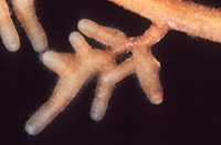
|
|
– Enlarged view – |
| • references | |
| Brand F (1988) Fagirhiza cystidiophora. In Agerer R (ed) Colour Atlas of Ectomycorrhizae, plate 15, Einhorn-Verlag, Schwäbisch Gmünd. Brand F, Agerer R (1988) Studien an Ektomykorrhizen XIII. Drei häufige Ektomykorrhizen der Buche (Fagus sylvatica L.). Charakterisierung und unsterile Kultivierung von Buchenektomykorrhizen. Sydowia 40: 1-37. |
|
| • length | |
| 0 mm | Lower value of unspecified range (could be µ-s.d., but not known) |
| 6 mm | Upper value of unspecified range (could be µ+s.d., but not known) |
| • ramification presence-type | |
| monopodial-pyramidal | |
| • tips {per 10 mm} number | |
| 20 | Lower value of unspecified range (could be µ-s.d., but not known) |
| 45 | Upper value of unspecified range (could be µ+s.d., but not known) |
| • ramification orders | |
| 0 | Lower value of unspecified range (could be µ-s.d., but not known) |
| 2 | Upper value of unspecified range (could be µ+s.d., but not known) |
| 3 | Maximum value |
| • main axis diameter | |
| 0.3 mm | Mean (= average) |
| 0.6 mm | Maximum value |
| • rhizomorphs as stout, short, conical structures presence-abundance | |
| absent | |
| • rhizomorphs as short mycorrhiza-like outgrowths with blunt tips presence | |
| absent | |
| • rhizomorphs presence | |
| absent | |
| • shape | |
| straight | |
| • shape {of distal end} | |
| not inflated, cylindric | |
| • length | |
| 0 mm | Lower value of unspecified range (could be µ-s.d., but not known) |
| 1.5 mm | Upper value of unspecified range (could be µ+s.d., but not known) |
| 3 mm | Maximum value |
| • diameter | |
| 0.2 mm | Lower value of unspecified range (could be µ-s.d., but not known) |
| 0.25 mm | Upper value of unspecified range (could be µ+s.d., but not known) |
| • colour | |
| yellow | |
| • very tip colour | |
| yellowish | |
| • older parts colour | |
| brown | |
| or | ochre, yellowish brown |
| • mantle {distinct} surface visibility | |
| present | |
| • mantle laticifers visibility | |
| absent | |
| • mantle dots presence-colour | |
| absent | |
| • mantle carbonizing presence | |
| absent | |
| • mantle surface {in general} habit | |
| not smooth | |
| • mantle surface {in detail} kind | |
| densely short-spiny | |
| • emanating hyphae presence | |
| present | |
| • emanating hyphae abundance | |
| infrequent | |
| • emanating hyphae distribution | |
| concentrated distally | |
| • presence | |
| absent | |
| • emanating elements presence-type | |
| cystidia | |
| • emanating elements cystidia location | |
| on outer mantle layer | |
| • presence | |
| absent | |
| • matrix presence | |
| present | |
| • matrix location | |
| outer mantle layer {apart from tip} | |
| • organisation | |
| pseudoparenchymatous | |
| • organisation {if pseudoparenchymatous} cell shape | |
| epidermoid, puzzle-like, jig-saw-shaped | |
| • mantle type | |
| epidermoid cells bearing a hyphal net (type Q) | |
| • hyphal net cells shape | |
| not specialized | |
| • hyphal net cells diameter | |
| 2 µm | Lower value of unspecified range (could be µ-s.d., but not known) |
| 7 µm | Upper value of unspecified range (could be µ+s.d., but not known) |
| • hyphal net cell walls habit | |
| distinct, rather thick | |
| • matrix kind | |
| not gelatinous | |
| • pores between cells presence | |
| absent | |
| • septa thickness {relative to cell walls} | |
| thinner than walls | |
| • septa clamps presence | |
| absent | |
| • cell pigment location-colour | |
| membranaceously yellowish | |
| • cell diameter | |
| 3 µm | Minimum value |
| 5 µm | Mean (= average) |
| • cell density | |
| 5 | Mean (= average) |
| • cell wall thickness | |
| 1.5 µm | Lower value of unspecified range (could be µ-s.d., but not known) |
| 3 µm | Upper value of unspecified range (could be µ+s.d., but not known) |
| • cell wall surface habit | |
| smooth | |
| • cell wall projections presence | |
| absent | |
| • drops of exuded pigment presence | |
| absent | |
| • organisation | |
| pseudoparenchymatous | |
| • cell pigment location-colour | |
| colourless | |
| • cell density | |
| 6 | Mean (= average) |
| • cell contents presence-kind | |
| absent | |
| • cell wall thickness | |
| 0.2 µm | Mean (= average) |
| • cell wall surface habit | |
| smooth | |
| • organisation | |
| plectenchymatous | |
| • hyphae arrangement | |
| without pattern | |
| • septa clamps presence | |
| absent | |
| • cell diameter | |
| 5 µm | Mean (= average) |
| • cell contents presence-kind | |
| absent | |
| • anatomy mantle outer mantle layer {of ectomycorrhizal tip} organisation | |
| like other parts of mantle | |
| or | pseudoparenchymatous |
| • anatomy mantle outer mantle layer {of ectomycorrhizal tip} cell density | |
| 7 | Mean (= average) |
| • mantle thickness {apart from tip} | |
| 15 µm | Minimum value |
| 20 µm | Lower value of unspecified range (could be µ-s.d., but not known) |
| 30 µm | Upper value of unspecified range (could be µ+s.d., but not known) |
| • mantle different layers presence | |
| discernable | |
| • outer mantle layer organisation | |
| plectenchymatous | |
| • middle mantle layer organisation | |
| pseudoparenchymatous | |
| • middle mantle layer hyphae tangentially length | |
| 5 µm | Lower value of unspecified range (could be µ-s.d., but not known) |
| 12 µm | Upper value of unspecified range (could be µ+s.d., but not known) |
| • middle mantle layer hyphae radially diameter | |
| 3 µm | Lower value of unspecified range (could be µ-s.d., but not known) |
| 7 µm | Upper value of unspecified range (could be µ+s.d., but not known) |
| • inner mantle layer organisation | |
| plectenchymatous | |
| • inner mantle layer hyphae radially diameter | |
| 3 µm | Lower value of unspecified range (could be µ-s.d., but not known) |
| 5 µm | Upper value of unspecified range (could be µ+s.d., but not known) |
| • presence | |
| absent | |
| • anatomy mantle longitudinal section cortical (epidermal) cells shape | |
| rectangular | |
| or | tangentially-oval to -elliptic or -cylindrical, and oriented obliquely |
| • anatomy mantle longitudinal section cortical (epidermal) cells radially diameter | |
| 0 µm | Lower value of unspecified range (could be µ-s.d., but not known) |
| 30 µm | Upper value of unspecified range (could be µ+s.d., but not known) |
| 35 µm | Maximum value |
| • mantle different layers presence | |
| discernible | |
| • outer mantle layer organisation | |
| plectenchymatous | |
| • middle mantle layer organisation | |
| pseudoparenchymatous | |
| • middle mantle layer hyphae tangentially length | |
| 5 µm | Lower value of unspecified range (could be µ-s.d., but not known) |
| 12 µm | Upper value of unspecified range (could be µ+s.d., but not known) |
| • middle mantle layer hyphae radially diameter | |
| 3 µm | Lower value of unspecified range (could be µ-s.d., but not known) |
| 7 µm | Upper value of unspecified range (could be µ+s.d., but not known) |
| • inner mantle layer organisation | |
| plectenchymatous | |
| • inner mantle layer hyphae radially diameter | |
| 3 µm | Lower value of unspecified range (could be µ-s.d., but not known) |
| 5 µm | Upper value of unspecified range (could be µ+s.d., but not known) |
| • presence | |
| absent | |
| • shape | |
| round | |
| or | radially-oval to -elliptic |
| • anatomy mantle cross-section cortical (epidermal) cells tangentially length | |
| 15 µm | Lower value of unspecified range (could be µ-s.d., but not known) |
| 25 µm | Upper value of unspecified range (could be µ+s.d., but not known) |
| • anatomy mantle cross-section cortical (epidermal) cells radially diameter | |
| 20 µm | Lower value of unspecified range (could be µ-s.d., but not known) |
| 30 µm | Upper value of unspecified range (could be µ+s.d., but not known) |
| • anatomy mantle cross-section hyphal cells around cortical (epidermal) cells shape | |
| cylindrical | |
| • anatomy mantle cross-section hyphal cells around cortical (epidermal) cells thickness | |
| 3.5 µm | Lower value of unspecified range (could be µ-s.d., but not known) |
| 4 µm | Upper value of unspecified range (could be µ+s.d., but not known) |
| 5 µm | Maximum value |
| • anatomy mantle cross-section hyphal rows around cortical (epidermal) cells number | |
| one | |
| • lobes width | |
| 2 µm | Lower value of unspecified range (could be µ-s.d., but not known) |
| 5 µm | Upper value of unspecified range (could be µ+s.d., but not known) |
| • structure {in plan view} | |
| of palmetti type | |
| • backwards-oriented clamps presence | |
| absent | |
| • anastomoses type | |
| closed by a simple septum, with a short bridge or bridge almost lacking (contact-septum) | |
| • anastomoses cell wall thickness {relative to remaining cell walls} | |
| as thick as | |
| • anastomoses anastomosal bridge thickness {relative to hyphae} | |
| thinner | |
| • type | |
| awl-shaped, bristle-like (type A) | |
| • ramification presence-position | |
| absent | |
| • septa presence | |
| present | |
| • septa kind | |
| simple | |
| • diameter {proximal} | |
| 4 µm | Lower value of unspecified range (could be µ-s.d., but not known) |
| 5 µm | Upper value of unspecified range (could be µ+s.d., but not known) |
| • length | |
| 80 µm | Lower value of unspecified range (could be µ-s.d., but not known) |
| 120 µm | Upper value of unspecified range (could be µ+s.d., but not known) |
| • cell wall thickness | |
| 0.5 µm | Lower value of unspecified range (could be µ-s.d., but not known) |
| 1 µm | Upper value of unspecified range (could be µ+s.d., but not known) |
| • cell wall thickness {relative to mantle cells} | |
| thinner | |
| • cell wall evenness | |
| even in thickness | |
| • surface habit | |
| smooth | |
| • contents presence | |
| absent | |
| • contents type | |
| absent | |
| • shape | |
| not striking | |
| • cell pigment location-colour | |
| absent | |
| • drops of exuded pigment presence | |
| absent | |
| • presence | |
| present | |
| • kind | |
| acute | |
| • clamps presence | |
| absent | |
| • clamps hole presence | |
| absent | |
| • clamps blister-like structure {at basis} presence | |
| absent | |
| • anatomy emanating elements emanating hyphae cell shape | |
| even | |
| • anatomy emanating elements emanating hyphae cell shape {at distal end} | |
| simple | |
| • anatomy emanating elements emanating hyphae cell diameter | |
| 4 µm | Minimum value |
| 4.5 µm | Mean (= average) |
| 5 µm | Maximum value |
| • anatomy emanating elements emanating hyphae cell length | |
| 20 µm | Lower value of unspecified range (could be µ-s.d., but not known) |
| 100 µm | Upper value of unspecified range (could be µ+s.d., but not known) |
| • anatomy emanating elements emanating hyphae cell wall surface habit | |
| smooth | |
| or | without lens-shaped appositions |
| or | without spindle-shaped appositions |
| • anatomy emanating elements emanating hyphae cell wall thickness | |
| 0.2 µm | Mean (= average) |
| • anatomy emanating elements emanating hyphae cell wall {apart from tip} evenness | |
| even in thickness | |
| • type | |
| lacking, only emanating hyphae present (type G) |
|
| • presence | |
| absent | |
| • {of ectomycorrhiza former} presence | |
| present | |
| • {of ectomycorrhiza former} abundance | |
| occasionally present | |
| • shape | |
| globular | |
| • diameter | |
| 5 µm | Lower value of unspecified range (could be µ-s.d., but not known) |
| 9 µm | Upper value of unspecified range (could be µ+s.d., but not known) |
| • number {per cell} | |
| 1 | Mean (= average) |
| 2 | Maximum value |
| • shape | |
| round | |
| • diameter | |
| 1.5 µm | Mean (= average) |
| 2 µm | Maximum value |
| • length | |
| 1.5 µm | Mean (= average) |
| 2 µm | Maximum value |
| • distance {between each other} | |
| 2 µm | Mean (= average) |
| 3 µm | Maximum value |
| • geographic occurrence continent | |
| Europe | |
| • plant family | |
| Fagaceae | |
| • plant genus | |
| Fagus | |
| • plant habitat kind | |
| forests, woods | |
| • public notes | |
| Mycorrhizal ends straw-coloured to golden-yellow, older parts golden-brown to honey-coloured; outer mantle layers in plan view with indivudual hyphae often still discernible; middle mantle layers in plan view with isodiametric to slightly elongated, partially rectangular, partly slightly epidermoid cells; cystidia with 1-3 simple septa; in cross-section Hartig net 1-2(-3) cortical cell layers deep; autofluorescence of mantle in section with UV-filter outer layers creme-white and inner layers slightly brownish orange, with blue-filter greenish yellow throughout; mantle in KOH light nut-brown, in brillant-cresyl-blue bluish, in toluidin-blue blue, in cotton-blue outer mantle cell walls distinctly blue, in ethanol slowly bleaching, in ruthenium-red outer mantle cell walls reddish, in Kongo-red red. | |