|
|
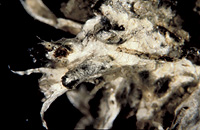
– Enlarged view – |
| • references | |
| Agerer R (1993) Ectomycorrhizae of Hydnellum peckii on Norway spruce and their chlamydospores. Mycologia 85: 74-83. Agerer R (1993) Hydnellum peckii. In Agerer R (ed) Colour Atlas of Ectomycorrhizae, plate 77, Einhorn-Verlag, Schwäbisch Gmünd. |
|
| • length | |
| 0 mm | Lower value of unspecified range (could be µ-s.d., but not known) |
| 20 mm | Upper value of unspecified range (could be µ+s.d., but not known) |
| • ramification presence-type | |
| monopodial-pyramidal | |
| • abundance | |
| nest-like, hyphal mats | |
| • main axis diameter | |
| 0.36 mm | Minimum value |
| 0.45 mm | Lower value of unspecified range (could be µ-s.d., but not known) |
| 0.7 mm | Upper value of unspecified range (could be µ+s.d., but not known) |
| 1.3 mm | Maximum value |
| • rhizomorphs as stout, short, conical structures presence-abundance | |
| absent | |
| • rhizomorphs as short mycorrhiza-like outgrowths with blunt tips presence | |
| absent | |
| • rhizomorphs presence | |
| present | |
| • rhizomorphs frequency | |
| abundant | |
| • exploration type | |
| medium distance mat | |
| • shape | |
| bent | |
| or | tortuous |
| • shape {of distal end} | |
| not inflated, cylindric | |
| or | tapering |
| • length | |
| 0 mm | Lower value of unspecified range (could be µ-s.d., but not known) |
| 2.3 mm | Upper value of unspecified range (could be µ+s.d., but not known) |
| 2.9 mm | Maximum value |
| • diameter | |
| 0.27 mm | Minimum value |
| 0.33 mm | Lower value of unspecified range (could be µ-s.d., but not known) |
| 0.39 mm | Upper value of unspecified range (could be µ+s.d., but not known) |
| 0.45 mm | Maximum value |
| • colour | |
| brown | |
| or | violet, dark reddish blue |
| • very tip colour | |
| violet, dark reddish blue | |
| or | white |
| • older parts colour | |
| black | |
| • mantle cortical cells visibility | |
| not visible | |
| • mantle {distinct} surface visibility | |
| present | |
| • mantle transparency | |
| not transparent | |
| • mantle laticifers visibility | |
| absent | |
| • mantle dots presence-colour | |
| yellowish or greenish | |
| • mantle carbonizing presence | |
| present | |
| • mantle surface {in general} habit | |
| silvery | |
| or | smooth |
| or | not smooth |
| • mantle surface {in detail} kind | |
| loosely stringy | |
| • emanating hyphae presence | |
| present | |
| • emanating hyphae abundance | |
| infrequent | |
| • diameter | |
| 0 mm | Lower value of unspecified range (could be µ-s.d., but not known) |
| 0.3 mm | Upper value of unspecified range (could be µ+s.d., but not known) |
| • cross-section shape | |
| round or roundish | |
| or | flat |
| • colour | |
| white | |
| • ramification kind-frequency | |
| repeatedly into smaller filaments | |
| • connection to mantle kind | |
| oblique | |
| • origin location | |
| not specific | |
| • margin habit | |
| smooth | |
| or | hairy |
| • dimorphism presence | |
| absent | |
| • presence | |
| absent | |
| • emanating elements presence-type | |
| rhizomorphs | |
| • blue granules presence | |
| present | |
| • blue granules location | |
| inner mantle layer | |
| • presence | |
| absent | |
| • matrix presence | |
| present | |
| • matrix location | |
| outer mantle layer {apart from tip} | |
| or | outer mantle layer {of ectomycorrhizal tip} |
| or | middle mantle layer |
| or | inner mantle layer |
| • organisation | |
| plectenchymatous | |
| • mantle type | |
| hyphae rather irregularly arranged and no special pattern discernible (type B) | |
| and | gelatinous matrix between the hyphae (type C) |
| • matrix kind | |
| gelatinous | |
| • hyphal system kind | |
| undifferentiated | |
| • septa thickness {relative to cell walls} | |
| thicker than walls | |
| • cell shape | |
| cylindric, not constricted at septa | |
| and | cylindric, constricted at septa |
| • cell pigment location-colour | |
| absent | |
| and | membranaceously yellowish |
| and | membranaceously greyish |
| and | plasmatically yellowish |
| • cell diameter | |
| 2 µm | Lower value of unspecified range (could be µ-s.d., but not known) |
| 3 µm | Upper value of unspecified range (could be µ+s.d., but not known) |
| • cell wall thickness | |
| 0.2 µm | Mean (= average) |
| • cell wall surface habit | |
| with many soil particles | |
| • drops of exuded pigment presence | |
| absent | |
| • organisation | |
| plectenchymatous | |
| • matrix kind | |
| not gelatinous | |
| • hyphae arrangement | |
| plectenchymatous, without pattern | |
| • cell pigment location-colour | |
| membranaceously brownish | |
| • cell diameter | |
| 1.5 µm | Minimum value |
| 2 µm | Lower value of unspecified range (could be µ-s.d., but not known) |
| 3 µm | Upper value of unspecified range (could be µ+s.d., but not known) |
| • cell wall thickness | |
| 0.2 µm | Mean (= average) |
| • cell wall surface habit | |
| smooth | |
| • organisation | |
| plectenchymatous with pseudoparenchymatous nests of cells | |
| • matrix kind | |
| gelatinous | |
| • hyphae arrangement | |
| ring-like | |
| • septa clamps presence | |
| present | |
| • cell diameter | |
| 1.5 µm | Minimum value |
| 2 µm | Lower value of unspecified range (could be µ-s.d., but not known) |
| 4 µm | Upper value of unspecified range (could be µ+s.d., but not known) |
| 6 µm | Maximum value |
| • cell blue granules location | |
| within cells | |
| • anatomy mantle outer mantle layer {of ectomycorrhizal tip} organisation | |
| like other parts of mantle | |
| • anatomy mantle outer mantle layer {of ectomycorrhizal tip} matrix kind | |
| gelatinous | |
| • anatomy mantle outer mantle layer {of ectomycorrhizal tip} hyphae diameter | |
| 1 µm | Lower value of unspecified range (could be µ-s.d., but not known) |
| 2 µm | Upper value of unspecified range (could be µ+s.d., but not known) |
| • mantle thickness {apart from tip} | |
| 15 µm | Minimum value |
| 20 µm | Lower value of unspecified range (could be µ-s.d., but not known) |
| 30 µm | Upper value of unspecified range (could be µ+s.d., but not known) |
| 40 µm | Maximum value |
| • mantle thickness {at ectomycorrhizal tip} | |
| 25 µm | Lower value of unspecified range (could be µ-s.d., but not known) |
| 40 µm | Upper value of unspecified range (could be µ+s.d., but not known) |
| • mantle different layers presence | |
| discernable | |
| • outer mantle layer organisation | |
| plectenchymatous | |
| • outer mantle layer hyphae tangentially length | |
| 2 µm | Lower value of unspecified range (could be µ-s.d., but not known) |
| 20 µm | Upper value of unspecified range (could be µ+s.d., but not known) |
| 40 µm | Maximum value |
| • outer mantle layer hyphae radially diameter | |
| 1.5 µm | Minimum value |
| 2 µm | Lower value of unspecified range (could be µ-s.d., but not known) |
| 4 µm | Upper value of unspecified range (could be µ+s.d., but not known) |
| 7.5 µm | Maximum value |
| • middle mantle layer organisation | |
| plectenchymatous | |
| • inner mantle layer organisation | |
| plectenchymatous | |
| • inner mantle layer hyphae tangentially length | |
| 2 µm | Lower value of unspecified range (could be µ-s.d., but not known) |
| 20 µm | Upper value of unspecified range (could be µ+s.d., but not known) |
| 60 µm | Maximum value |
| • inner mantle layer hyphae radially diameter | |
| 2 µm | Minimum value |
| 3 µm | Lower value of unspecified range (could be µ-s.d., but not known) |
| 5 µm | Upper value of unspecified range (could be µ+s.d., but not known) |
| • presence | |
| present | |
| • shape | |
| tangentially-oval, -elliptic or -cylindrical, and oriented in parallel to root axis | |
| • tangentially length | |
| 25 µm | Minimum value |
| 40 µm | Lower value of unspecified range (could be µ-s.d., but not known) |
| 145 µm | Upper value of unspecified range (could be µ+s.d., but not known) |
| 165 µm | Maximum value |
| • radially diameter | |
| 5 µm | Lower value of unspecified range (could be µ-s.d., but not known) |
| 25 µm | Upper value of unspecified range (could be µ+s.d., but not known) |
| 30 µm | Maximum value |
| • mean tangenial length TCt | |
| 52 µm | Lower value of unspecified range (could be µ-s.d., but not known) |
| 97.5 µm | Upper value of unspecified range (could be µ+s.d., but not known) |
| • mean shape-ratio TCq | |
| 3 | Lower value of unspecified range (could be µ-s.d., but not known) |
| 7.1 | Upper value of unspecified range (could be µ+s.d., but not known) |
| • anatomy mantle longitudinal section cortical (epidermal) cells shape | |
| tangentially-oval to -elliptic or -cylindrical, and oriented obliquely | |
| • anatomy mantle longitudinal section cortical (epidermal) cells tangentially length | |
| 13 µm | Minimum value |
| 20 µm | Lower value of unspecified range (could be µ-s.d., but not known) |
| 130 µm | Upper value of unspecified range (could be µ+s.d., but not known) |
| 157 µm | Maximum value |
| • anatomy mantle longitudinal section cortical (epidermal) cells radially diameter | |
| 12 µm | Lower value of unspecified range (could be µ-s.d., but not known) |
| 40 µm | Upper value of unspecified range (could be µ+s.d., but not known) |
| 45 µm | Maximum value |
| • anatomy mantle longitudinal section cortical (epidermal) cells mean tangential length CCt (ECt) | |
| 24.5 µm | Lower value of unspecified range (could be µ-s.d., but not known) |
| 91.6 µm | Upper value of unspecified range (could be µ+s.d., but not known) |
| • anatomy mantle longitudinal section cortical (epidermal) cells mean shape-ratio CCq (ECq) | |
| 1.4 | Lower value of unspecified range (could be µ-s.d., but not known) |
| 4.2 | Upper value of unspecified range (could be µ+s.d., but not known) |
| • presence | |
| present | |
| • kind | |
| protruding towards endodermis | |
| • structure {in plan view} | |
| of palmetti type | |
| • lobes width | |
| 1 µm | Minimum value |
| 2 µm | Lower value of unspecified range (could be µ-s.d., but not known) |
| 3 µm | Upper value of unspecified range (could be µ+s.d., but not known) |
| 4 µm | Maximum value |
| • mantle different layers presence | |
| not discernible | |
| • outer mantle layer organisation | |
| plectenchymatous | |
| • middle mantle layer organisation | |
| plectenchymatous | |
| • inner mantle layer organisation | |
| plectenchymatous | |
| • presence | |
| present | |
| • rows number | |
| 1 | Mean (= average) |
| 2 | Maximum value |
| • shape | |
| tangentially-oval to tangentially-elliptic | |
| • tangentially length | |
| 11 µm | Minimum value |
| 20 µm | Lower value of unspecified range (could be µ-s.d., but not known) |
| 45 µm | Upper value of unspecified range (could be µ+s.d., but not known) |
| 65 µm | Maximum value |
| • radially diameter | |
| 5 µm | Lower value of unspecified range (could be µ-s.d., but not known) |
| 18 µm | Upper value of unspecified range (could be µ+s.d., but not known) |
| 28 µm | Maximum value |
| • mean tangential length TCt | |
| 29.5 µm | Lower value of unspecified range (could be µ-s.d., but not known) |
| 37.5 µm | Upper value of unspecified range (could be µ+s.d., but not known) |
| • mean shape-ratio TCq | |
| 2.7 | Lower value of unspecified range (could be µ-s.d., but not known) |
| 3.9 | Upper value of unspecified range (could be µ+s.d., but not known) |
| • anatomy mantle cross-section cortical (epidermal) cells shape | |
| radially-oval to -elliptic | |
| or | tangentially-oval to tangentially-elliptic |
| • anatomy mantle cross-section cortical (epidermal) cells tangentially length | |
| 6 µm | Minimum value |
| 15 µm | Lower value of unspecified range (could be µ-s.d., but not known) |
| 36 µm | Upper value of unspecified range (could be µ+s.d., but not known) |
| 46 µm | Maximum value |
| • anatomy mantle cross-section cortical (epidermal) cells radially diameter | |
| 10 µm | Minimum value |
| 15 µm | Lower value of unspecified range (could be µ-s.d., but not known) |
| 32 µm | Upper value of unspecified range (could be µ+s.d., but not known) |
| 42 µm | Maximum value |
| • anatomy mantle cross-section cortical (epidermal) cells mean tangential length CCt | |
| 22.3 µm | Lower value of unspecified range (could be µ-s.d., but not known) |
| 26.8 µm | Upper value of unspecified range (could be µ+s.d., but not known) |
| • anatomy mantle cross-section cortical (epidermal) cells mean shape-ratio CCq | |
| 1.1 | Lower value of unspecified range (could be µ-s.d., but not known) |
| 1.4 | Upper value of unspecified range (could be µ+s.d., but not known) |
| • presence | |
| present | |
| • kind | |
| protruding towards endodermis | |
| • anatomy mantle cross-section hyphal cells around tannin cells shape | |
| roundish | |
| or | cylindrical |
| • anatomy mantle cross-section hyphal cells around tannin cells thickness | |
| 3 µm | Lower value of unspecified range (could be µ-s.d., but not known) |
| 4 µm | Upper value of unspecified range (could be µ+s.d., but not known) |
| • anatomy mantle cross-section hyphal rows around tannin cells number | |
| one | |
| • anatomy mantle cross-section hyphal cells around cortical (epidermal) cells shape | |
| roundish | |
| or | cylindrical |
| • anatomy mantle cross-section hyphal cells around cortical (epidermal) cells thickness | |
| 2 µm | Lower value of unspecified range (could be µ-s.d., but not known) |
| 3 µm | Upper value of unspecified range (could be µ+s.d., but not known) |
| 4 µm | Maximum value |
| • anatomy mantle cross-section hyphal rows around cortical (epidermal) cells number | |
| one | |
| • septal pores configuration | |
| globular thickenings | |
| • anastomoses type | |
| open, with a short bridge or bridge almost lacking | |
| • anastomoses cell wall thickness {relative to remaining cell walls} | |
| as thick as | |
| • anastomoses location | |
| close to hyphal tips | |
| • shape | |
| not striking | |
| • cell pigment location-colour | |
| absent | |
| or | membranaceously yellowish |
| • drops of exuded pigment presence | |
| absent | |
| • clamps presence | |
| present | |
| • clamps outline {in lateral view} | |
| less than a semicircle | |
| or | not constricted at contact point to subtending hyphal cell |
| • clamps width {relative to hypha in lateral view} | |
| thinner than | |
| • clamps hole presence | |
| absent | |
| • clamps blister-like structure {at basis} presence | |
| absent | |
| • presence | |
| present | |
| • abundance | |
| abundant | |
| • distribution | |
| evenly distributed | |
| • anatomy emanating elements emanating hyphae cell shape {at distal end} | |
| simple | |
| • anatomy emanating elements emanating hyphae cell diameter | |
| 1.5 µm | Lower value of unspecified range (could be µ-s.d., but not known) |
| 2 µm | Upper value of unspecified range (could be µ+s.d., but not known) |
| • anatomy emanating elements emanating hyphae cell wall surface habit | |
| smooth | |
| or | without lens-shaped appositions |
| or | without spindle-shaped appositions |
| or | with soil particles |
| • type | |
| differentiated; some hyphae very thick, which appear randomly distributed, pores of septa somtimes enlarged (type D) |
|
| • nodia presence | |
| absent | |
| • internal nodia presence | |
| absent | |
| • gelatinous matrix presence | |
| present | |
| • gelatinized hyphae presence | |
| absent | |
| • conical young side-branches presence | |
| absent | |
| • a "ball" of intertwined, ramified, thin hyphae presence | |
| absent | |
| • ampullate, trumpet-like inflated presence | |
| present | |
| • ampullate inflations thickness | |
| 6 µm | Lower value of unspecified range (could be µ-s.d., but not known) |
| 10 µm | Upper value of unspecified range (could be µ+s.d., but not known) |
| • anatomy emanating elements rhizomorphs hyphae ampullate, trumpet-like inflated presence | |
| present | |
| • anatomy emanating elements rhizomorphs hyphae ampullate inflations thickness | |
| 6 µm | Lower value of unspecified range (could be µ-s.d., but not known) |
| 10 µm | Upper value of unspecified range (could be µ+s.d., but not known) |
| • anatomy emanating elements rhizomorphs hyphae ampullate, trumpet-like inflated presence | |
| present | |
| • anatomy emanating elements rhizomorphs hyphae ampullate inflations thickness | |
| 6 µm | Lower value of unspecified range (could be µ-s.d., but not known) |
| 10 µm | Upper value of unspecified range (could be µ+s.d., but not known) |
| • anatomy emanating elements rhizomorphs hyphae ampullate, trumpet-like inflated presence | |
| present | |
| • anatomy emanating elements rhizomorphs hyphae ampullate inflations thickness | |
| 6 µm | Lower value of unspecified range (could be µ-s.d., but not known) |
| 10 µm | Upper value of unspecified range (could be µ+s.d., but not known) |
| • anatomy emanating elements rhizomorphs hyphae ampullate, trumpet-like inflated presence | |
| present | |
| • anatomy emanating elements rhizomorphs hyphae ampullate inflations thickness | |
| 6 µm | Lower value of unspecified range (could be µ-s.d., but not known) |
| 10 µm | Upper value of unspecified range (could be µ+s.d., but not known) |
| • presence | |
| present | |
| • type | |
| with radially splitting walls | |
| • diameter | |
| 9 µm | Minimum value |
| 11 µm | Lower value of unspecified range (could be µ-s.d., but not known) |
| 13 µm | Upper value of unspecified range (could be µ+s.d., but not known) |
| 15 µm | Maximum value |
| • length | |
| 9 µm | Minimum value |
| 11 µm | Lower value of unspecified range (could be µ-s.d., but not known) |
| 13 µm | Upper value of unspecified range (could be µ+s.d., but not known) |
| 15 µm | Maximum value |
| • {of ectomycorrhiza former} presence | |
| absent | |
| • {of foreign origin} presence | |
| absent | |
| • number {per cell} | |
| 2 | Mean (= average) |
| 3 | Maximum value |
| • shape | |
| round | |
| • diameter | |
| 1.5 µm | Mean (= average) |
| 2 µm | Maximum value |
| • length | |
| 1.5 µm | Mean (= average) |
| 2 µm | Maximum value |
| • distance {between each other} | |
| 0 µm | Lower value of unspecified range (could be µ-s.d., but not known) |
| 6 µm | Upper value of unspecified range (could be µ+s.d., but not known) |
| • presence | |
| present | |
| • frequency | |
| in few cells | |
| • arrangement | |
| solitary | |
| • diameter | |
| 0 µm | Lower value of unspecified range (could be µ-s.d., but not known) |
| 0.5 µm | Upper value of unspecified range (could be µ+s.d., but not known) |
| • substrate | |
| in organically enriched mineral horizon | |
| or | in mineral soil |
| • geographic occurrence continent | |
| Europe | |
| • knowledge about association with foreign fruitbodies presence | |
| unknown | |
| • plant family | |
| Pinaceae | |
| • plant genus | |
| Picea | |
| • plant habitat kind | |
| forests, woods | |
| • family | |
| Bankeraceae ss. Stalpers | |
| • fruitbodies growth habit | |
| epigeous | |
| or | pileate-hydnoid |
| • public notes | |
| Middle and inner mantle layers in plan view membranaceously greyish; autofluorescence of mantle in section with UV-filter weakly greenish blue, with blue-filter weakly yellowish, with green-filter slightly reddish; mantle in KOH reddish brown, in lactic acid distinctly yellowish brown and blue granules appear at least after longer storage, in brillant-cresyl-blue bluish green after rinsing with water, in cotton-blue walls and cytoplsm slightly blue, in anilin walls pinkish. | |