|
|
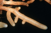
– Enlarged view – |
| • references | |
| Luppi AM, Gautero C (1967) Ricerche sulle micorrize di Quercus robur, Q. petraea e Q. pubescens in Piemonte. Allionia 13: 129-148. Palfner G (1998) Lactarius chrysorrheus. In Agerer R (ed) Colour Atlas of Ectomycorrhizae, plate 121, Einhorn-Verlag, Schwäbisch Gmünd. Palfner G, Agerer R (1996) Die Ektomykorrhizen von Lactarius chrysorrheus und L. serifluus an Quercus robur. Sendtnera 3: 119-136. |
|
| • length | |
| 0 mm | Lower value of unspecified range (could be µ-s.d., but not known) |
| 23 mm | Upper value of unspecified range (could be µ+s.d., but not known) |
| • ramification presence-type | |
| monopodial-pinnate | |
| or | monopodial-pyramidal |
| • ramification orders | |
| 0 | Lower value of unspecified range (could be µ-s.d., but not known) |
| 3 | Upper value of unspecified range (could be µ+s.d., but not known) |
| • abundance | |
| abundant, dense | |
| • main axis diameter | |
| 0.3 mm | Lower value of unspecified range (could be µ-s.d., but not known) |
| 0.5 mm | Upper value of unspecified range (could be µ+s.d., but not known) |
| • rhizomorphs as stout, short, conical structures presence-abundance | |
| absent | |
| • rhizomorphs as short mycorrhiza-like outgrowths with blunt tips presence | |
| absent | |
| • rhizomorphs presence | |
| present | |
| • rhizomorphs frequency | |
| abundant | |
| • shape | |
| straight | |
| or | bent |
| • shape {of distal end} | |
| not inflated, cylindric | |
| • length | |
| 0 mm | Lower value of unspecified range (could be µ-s.d., but not known) |
| 2 mm | Upper value of unspecified range (could be µ+s.d., but not known) |
| 3 mm | Maximum value |
| • diameter | |
| 0.3 mm | Mean (= average) |
| 0.4 mm | Maximum value |
| • colour | |
| ochre, yellowish brown | |
| or | white |
| • older parts colour | |
| dark brown | |
| • mantle cortical cells visibility | |
| not visible | |
| • mantle {distinct} surface visibility | |
| present | |
| • mantle transparency | |
| not transparent | |
| • mantle laticifers visibility | |
| present | |
| • mantle secreted latex {after scratching mantle} presence | |
| present | |
| • mantle secreted latex {after scratching mantle} colour-habit | |
| white, remaining white after some minutes | |
| • mantle dots presence-colour | |
| absent | |
| • mantle carbonizing presence | |
| absent | |
| • mantle surface {in general} habit | |
| shiny | |
| or | smooth |
| • emanating hyphae presence | |
| present | |
| • emanating hyphae abundance | |
| infrequent | |
| • diameter | |
| 0.007 mm | Lower value of unspecified range (could be µ-s.d., but not known) |
| 0.19 mm | Upper value of unspecified range (could be µ+s.d., but not known) |
| • cross-section shape | |
| round or roundish | |
| • colour | |
| concolourous to mantle | |
| or | brown |
| or | orange |
| or | whitish |
| • connection to mantle kind | |
| oblique | |
| • margin habit | |
| smooth | |
| • dimorphism presence | |
| absent | |
| • presence | |
| absent | |
| • emanating elements presence-type | |
| rhizomorphs | |
| • blue granules presence | |
| absent | |
| • presence | |
| absent | |
| or | present |
| • location | |
| middle mantle layer | |
| or | inner mantle layer |
| or | rhizomorphs |
| • shape | |
| straight and even | |
| or | worm-like, uneven |
| • ramification presence-abundance | |
| abundant | |
| • cell diameter | |
| 5 µm | Lower value of unspecified range (could be µ-s.d., but not known) |
| 8 µm | Upper value of unspecified range (could be µ+s.d., but not known) |
| 10 µm | Maximum value |
| • matrix presence | |
| present | |
| • matrix location | |
| outer mantle layer {apart from tip} | |
| or | outer mantle layer {of ectomycorrhizal tip} |
| or | middle mantle layer |
| • organisation | |
| pseudoparenchymatous | |
| • organisation {if pseudoparenchymatous} cell shape | |
| epidermoid, puzzle-like, jig-saw-shaped | |
| • mantle type | |
| epidermoid cells bearing a hyphal net (type Q) | |
| • hyphal net cells shape | |
| triangular with obtuse ends | |
| and | with roundish lobes |
| • hyphal net cells diameter | |
| 8 µm | Minimum value |
| 14 µm | Mean (= average) |
| 18 µm | Maximum value |
| • hyphal net cell walls habit | |
| faint, delicate, inconspicuous, rather thin | |
| • matrix kind | |
| not gelatinous | |
| • pores between cells presence | |
| absent | |
| • hyphae hyphal junctions angle {between} | |
| ca. 120° | |
| • septa clamps presence | |
| absent | |
| • cell diameter | |
| 5 µm | Minimum value |
| 11 µm | Mean (= average) |
| 16 µm | Maximum value |
| • cell length | |
| 5 µm | Minimum value |
| 10 µm | Mean (= average) |
| 20 µm | Maximum value |
| • cell density | |
| 6 | Minimum value |
| 9 | Mean (= average) |
| 11 | Maximum value |
| • cell wall thickness | |
| 0.5 µm | Lower value of unspecified range (could be µ-s.d., but not known) |
| 1 µm | Upper value of unspecified range (could be µ+s.d., but not known) |
| • cell wall surface habit | |
| smooth | |
| • cell wall projections presence | |
| present | |
| • cell wall projections abundance | |
| abundant | |
| • cell wall projections shape | |
| even in thickness | |
| and | thicker at distal end |
| • drops of exuded pigment presence | |
| absent | |
| • organisation | |
| plectenchymatous | |
| • matrix kind | |
| not gelatinous | |
| • hyphae arrangement | |
| plectenchymatous, without pattern | |
| • cell diameter | |
| 3 µm | Minimum value |
| 4 µm | Mean (= average) |
| 5 µm | Maximum value |
| • cell wall thickness | |
| 0.5 µm | Mean (= average) |
| • cell wall surface habit | |
| smooth | |
| • organisation | |
| plectenchymatous with pseudoparenchymatous nests of cells | |
| • hyphae arrangement | |
| ring-like | |
| • septa clamps presence | |
| absent | |
| • cell diameter | |
| 2 µm | Minimum value |
| 4 µm | Mean (= average) |
| 5 µm | Maximum value |
| • anatomy mantle outer mantle layer {of ectomycorrhizal tip} organisation | |
| pseudoparenchymatous | |
| • anatomy mantle outer mantle layer {of ectomycorrhizal tip} matrix kind | |
| not gelatinous | |
| • anatomy mantle outer mantle layer {of ectomycorrhizal tip} hyphae diameter | |
| 2 µm | Minimum value |
| 5 µm | Mean (= average) |
| 7 µm | Maximum value |
| • anatomy mantle outer mantle layer {of ectomycorrhizal tip} cell density | |
| 9 | Minimum value |
| 14 | Mean (= average) |
| 23 | Maximum value |
| • mantle thickness {apart from tip} | |
| 10 µm | Minimum value |
| 20 µm | Lower value of unspecified range (could be µ-s.d., but not known) |
| 30 µm | Upper value of unspecified range (could be µ+s.d., but not known) |
| • mantle different layers presence | |
| discernable | |
| • outer mantle layer organisation | |
| pseudoparenchymatous | |
| • outer mantle layer hyphae tangentially length | |
| 5 µm | Minimum value |
| 8 µm | Mean (= average) |
| 12 µm | Maximum value |
| • outer mantle layer hyphae radially diameter | |
| 1 µm | Minimum value |
| 2 µm | Mean (= average) |
| 3 µm | Maximum value |
| • middle mantle layer organisation | |
| plectenchymatous | |
| • middle mantle layer hyphae tangentially length | |
| 4 µm | Minimum value |
| 5 µm | Mean (= average) |
| 7 µm | Maximum value |
| • middle mantle layer hyphae radially diameter | |
| 2 µm | Minimum value |
| 3 µm | Mean (= average) |
| 4 µm | Maximum value |
| • inner mantle layer organisation | |
| plectenchymatous | |
| • inner mantle layer hyphae tangentially length | |
| 3 µm | Minimum value |
| 6 µm | Mean (= average) |
| 12 µm | Maximum value |
| • inner mantle layer hyphae radially diameter | |
| 2 µm | Minimum value |
| 4 µm | Mean (= average) |
| 5 µm | Maximum value |
| • unlayered mantle hyphae radially diameter | |
| 3.5 µm | Lower value of unspecified range (could be µ-s.d., but not known) |
| 8.5 µm | Upper value of unspecified range (could be µ+s.d., but not known) |
| • laticifers diameter | |
| 4 µm | Minimum value |
| 7 µm | Mean (= average) |
| 11 µm | Maximum value |
| • presence | |
| absent | |
| • anatomy mantle longitudinal section cortical (epidermal) cells shape | |
| radially-oval to -elliptic, oriented obliquely | |
| • anatomy mantle longitudinal section cortical (epidermal) cells tangentially length | |
| 15 µm | Minimum value |
| 18 µm | Mean (= average) |
| 27 µm | Maximum value |
| • anatomy mantle longitudinal section cortical (epidermal) cells radially diameter | |
| 28 µm | Minimum value |
| 40 µm | Mean (= average) |
| 50 µm | Maximum value |
| • anatomy mantle longitudinal section cortical (epidermal) cells mean shape-ratio CCq (ECq) | |
| 0.45 | Mean (= average) |
| • presence | |
| present | |
| • kind | |
| periepidermal | |
| or | paraepidermal |
| • structure {in plan view} | |
| of palmetti type | |
| • lobes width | |
| 2 µm | Minimum value |
| 3 µm | Mean (= average) |
| 5 µm | Maximum value |
| • mantle different layers presence | |
| discernible | |
| • outer mantle layer organisation | |
| pseudoparenchymatous | |
| • outer mantle layer hyphae tangentially length | |
| 5 µm | Minimum value |
| 8 µm | Mean (= average) |
| 12 µm | Maximum value |
| • outer mantle layer hyphae radially diameter | |
| 1 µm | Minimum value |
| 2 µm | Mean (= average) |
| 3 µm | Maximum value |
| • middle mantle layer organisation | |
| plectenchymatous | |
| • middle mantle layer hyphae tangentially length | |
| 4 µm | Minimum value |
| 5 µm | Mean (= average) |
| 7 µm | Maximum value |
| • middle mantle layer hyphae radially diameter | |
| 2 µm | Minimum value |
| 3 µm | Mean (= average) |
| 4 µm | Maximum value |
| • inner mantle layer organisation | |
| plectenchymatous | |
| • inner mantle layer hyphae tangentially length | |
| 3 µm | Minimum value |
| 6 µm | Mean (= average) |
| 12 µm | Maximum value |
| • inner mantle layer hyphae radially diameter | |
| 2 µm | Minimum value |
| 4 µm | Mean (= average) |
| 5 µm | Maximum value |
| • laticifers diameter | |
| 4 µm | Minimum value |
| 7 µm | Mean (= average) |
| 11 µm | Maximum value |
| • presence | |
| absent | |
| • anatomy mantle cross-section cortical (epidermal) cells shape | |
| round | |
| • anatomy mantle cross-section cortical (epidermal) cells tangentially length | |
| 11 µm | Minimum value |
| 15 µm | Mean (= average) |
| 20 µm | Maximum value |
| • anatomy mantle cross-section cortical (epidermal) cells radially diameter | |
| 7 µm | Minimum value |
| 12 µm | Mean (= average) |
| 20 µm | Maximum value |
| • anatomy mantle cross-section cortical (epidermal) cells mean shape-ratio CCq | |
| 1.25 | Mean (= average) |
| • anatomy mantle cross-section hyphal rows around tannin cells number | |
| one | |
| • anatomy mantle cross-section hyphal cells around cortical (epidermal) cells thickness | |
| 2 µm | Mean (= average) |
| 3 µm | Maximum value |
| • intrahyphal hyphae presence | |
| absent | |
| • septal pores configuration | |
| globular thickenings | |
| • backwards-oriented ramifications presence | |
| present | |
| • backwards-oriented clamps presence | |
| absent | |
| • clamps presence | |
| absent | |
| • type | |
| undifferentiated; margins rather smooth; hyphae compactly arranged and of uniform diameter (type B) |
|
| or | slightly differentiated; central hyphae somewhat enlarged (type C) |
| • nodia presence | |
| absent | |
| • internal nodia presence | |
| absent | |
| • gelatinous matrix presence | |
| absent | |
| • gelatinized hyphae presence | |
| absent | |
| • cup-like structures on surface presence | |
| absent | |
| • a "ball" of intertwined, ramified, thin hyphae presence | |
| absent | |
| • ampullate, trumpet-like inflated presence | |
| absent | |
| • anatomy emanating elements rhizomorphs hyphae ampullate, trumpet-like inflated presence | |
| absent | |
| • anatomy emanating elements rhizomorphs hyphae ampullate, trumpet-like inflated presence | |
| absent | |
| • anatomy emanating elements rhizomorphs hyphae ampullate, trumpet-like inflated presence | |
| absent | |
| • anatomy emanating elements rhizomorphs hyphae ampullate, trumpet-like inflated presence | |
| absent | |
| • presence | |
| absent | |
| • {of ectomycorrhiza former} presence | |
| absent | |
| • number {per cell} | |
| 2 | Mean (= average) |
| • shape | |
| round | |
| • diameter | |
| 0.8 µm | Minimum value |
| 1.2 µm | Mean (= average) |
| 1.6 µm | Maximum value |
| • length | |
| 0.8 µm | Minimum value |
| 1.2 µm | Mean (= average) |
| 1.6 µm | Maximum value |
| • distance {between each other} | |
| 0 µm | Lower value of unspecified range (could be µ-s.d., but not known) |
| 4 µm | Upper value of unspecified range (could be µ+s.d., but not known) |
| • substrate | |
| in organic layer | |
| • geographic occurrence continent | |
| Europe | |
| • knowledge about association with foreign fruitbodies presence | |
| unknown | |
| • plant family | |
| Fagaceae | |
| • plant genus | |
| Quercus | |
| • plant habitat kind | |
| forests, woods | |
| • family | |
| Russulaceae | |
| • subgenus-section | |
| Lactarius sect. Lactarius | |
| • fruitbodies growth habit | |
| epigeous | |
| or | pileate-lamellate |
| • public notes | |
| Mycorrhizal ends at most slightly bent, whitish beige to ochre, mostly with a tinge of flesh-colour, older parts dark brown with a tinge of flesh-colour; rhizomorphs often coloured like the mantle, on older mycorrhizae brownish orange; in cross-section Hartig net 2-2.5 cortical cell layers deep; autofluorescence of mantle with UV-filter showing an outer stripe of distinctly lighter colour than layers below; mantle in sulfo-vanillin with dark violet laticifers, mantle in KOH slightly orange, in guaiac slightly grey, in brillant-cresyl-blue margin of mantle piece slightly violet, in phenole laticifers slightly yellowish, in acid fuchsin mantle rosy to red, laticifers more distinctly coloured, in ruthenium-red margin of mantle piece slightly reddish. | |