|
|
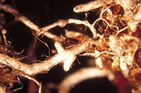
– Enlarged view – |
| • references | |
| Agerer R (1988) Studies on ectomycorrhizae XVII. The ontogeny of the ectomycorrhizal rhizomorphs of Paxillus involutus and Thelephora terrestris (Basidiomycetes). Nova Hedwigia 47: 311-334. Agerer R, Gronbachs E (1989) Paxillus involutus. In Agerer R (ed) Colour Atlas of Ectomycorrhizae, plate 27, Einhorn-Verlag, Schwäbisch Gmünd. Franz F, Acker G (1995) Rhizomorphs of Picea abies ectomycorrhizae: Ultrastructural aspects and element analysis (EELS and ESI) on hyphal inclusions. Nova Hedwigia 60(1-2): 253-267. Gronbach E (1988) Charakterisierung und Identifizierung von Ektomykorrhizen in einem Fichtenbestand mit Untersuchungen zur Merkmalsvariabilität in sauer beregneten Flächen. Bibliotheca Mycologica 125: 1-216. Ingleby K, Mason PA, Last FT, Fleming LV (1990) Identification of ectomycorrhizas. ITE Research Publication no. 5. HMSO, London. |
|
| • length | |
| 2 mm | Lower value of unspecified range (could be µ-s.d., but not known) |
| 13 mm | Upper value of unspecified range (could be µ+s.d., but not known) |
| 16 mm | Maximum value |
| • ramification presence-type | |
| monopodial-pinnate | |
| or | monopodial-pyramidal |
| • ramification orders | |
| 0 | Lower value of unspecified range (could be µ-s.d., but not known) |
| 1 | Upper value of unspecified range (could be µ+s.d., but not known) |
| • abundance | |
| solitary or in small numbers | |
| or | abundant, dense |
| • main axis diameter | |
| 0.4 mm | Lower value of unspecified range (could be µ-s.d., but not known) |
| 0.5 mm | Upper value of unspecified range (could be µ+s.d., but not known) |
| 0.6 mm | Maximum value |
| • rhizomorphs as stout, short, conical structures presence-abundance | |
| absent | |
| • rhizomorphs as short mycorrhiza-like outgrowths with blunt tips presence | |
| absent | |
| • rhizomorphs presence | |
| present | |
| • rhizomorphs frequency | |
| abundant | |
| • exploration type | |
| long distance | |
| • shape | |
| straight | |
| or | tortuous |
| • shape {of distal end} | |
| not inflated, cylindric | |
| • length | |
| 0 mm | Lower value of unspecified range (could be µ-s.d., but not known) |
| 3 mm | Upper value of unspecified range (could be µ+s.d., but not known) |
| • diameter | |
| 0.3 mm | Mean (= average) |
| 0.4 mm | Maximum value |
| • colour | |
| brown | |
| or | ochre, yellowish brown |
| or | white |
| • very tip colour | |
| whitish | |
| • older parts colour | |
| dark brown | |
| or | brown |
| • mantle cortical cells visibility | |
| not visible | |
| • mantle {distinct} surface visibility | |
| present | |
| • mantle transparency | |
| not transparent | |
| • mantle laticifers visibility | |
| absent | |
| • mantle dots presence-colour | |
| absent | |
| • mantle carbonizing presence | |
| absent | |
| • mantle surface {in general} habit | |
| silvery | |
| • mantle surface {in detail} kind | |
| densely stringy | |
| or | loosely stringy |
| • emanating hyphae presence | |
| present | |
| • emanating hyphae abundance | |
| infrequent | |
| • diameter | |
| 0.15 mm | Lower value of unspecified range (could be µ-s.d., but not known) |
| 0.8 mm | Upper value of unspecified range (could be µ+s.d., but not known) |
| • cross-section shape | |
| round or roundish | |
| • colour | |
| brown | |
| or | brownish |
| • ramification kind-frequency | |
| frequently, at restricted points | |
| or | infrequently, at restricted points |
| • connection to mantle kind | |
| distinct | |
| • margin habit | |
| smooth | |
| • dimorphism presence | |
| absent | |
| • presence | |
| present | |
| • abundance | |
| infrequent | |
| • shape | |
| globular | |
| • diameter | |
| 0.8 mm | Mean (= average) |
| • colour | |
| brown | |
| • formation location | |
| laterally on rhizomorphs | |
| • emanating elements presence-type | |
| rhizomorphs | |
| or | cystidia |
| • emanating elements cystidia location | |
| on outer mantle layer | |
| • blue granules presence | |
| absent | |
| • presence | |
| absent | |
| • matrix presence | |
| present | |
| • matrix location | |
| outer mantle layer {apart from tip} | |
| or | outer mantle layer {of ectomycorrhizal tip} |
| or | middle mantle layer |
| • organisation | |
| plectenchymatous | |
| • mantle type | |
| hyphae rather irregularly arranged and no special pattern discernible (type B) | |
| • matrix kind | |
| not gelatinous | |
| • hyphal system kind | |
| undifferentiated | |
| • hyphae hyphal junctions angle {between} | |
| ca. 45° and less | |
| • septa thickness {relative to cell walls} | |
| as thick as walls | |
| • cell shape | |
| cylindric, not constricted at septa | |
| • cell pigment location-colour | |
| absent | |
| • cell diameter | |
| 3 µm | Minimum value |
| 4 µm | Lower value of unspecified range (could be µ-s.d., but not known) |
| 5.5 µm | Upper value of unspecified range (could be µ+s.d., but not known) |
| 7 µm | Maximum value |
| • cell length | |
| 6 µm | Lower value of unspecified range (could be µ-s.d., but not known) |
| 60 µm | Upper value of unspecified range (could be µ+s.d., but not known) |
| • cell wall thickness | |
| 0.2 µm | Mean (= average) |
| • cell wall surface habit | |
| smooth | |
| • drops of exuded pigment presence | |
| absent | |
| • organisation | |
| plectenchymatous | |
| • matrix kind | |
| not gelatinous | |
| • hyphae arrangement | |
| plectenchymatous, without pattern | |
| • cell pigment location-colour | |
| colourless | |
| • cell diameter | |
| 3 µm | Minimum value |
| 4 µm | Lower value of unspecified range (could be µ-s.d., but not known) |
| 5.5 µm | Upper value of unspecified range (could be µ+s.d., but not known) |
| 6 µm | Maximum value |
| • cell length | |
| 6 µm | Lower value of unspecified range (could be µ-s.d., but not known) |
| 60 µm | Upper value of unspecified range (could be µ+s.d., but not known) |
| • cell wall thickness | |
| 0.2 µm | Mean (= average) |
| • cell wall surface habit | |
| smooth | |
| • organisation | |
| plectenchymatous | |
| • hyphae arrangement | |
| without pattern | |
| • septa clamps presence | |
| present | |
| • cell pigment location-colour | |
| absent | |
| • cell diameter | |
| 3 µm | Minimum value |
| 4 µm | Lower value of unspecified range (could be µ-s.d., but not known) |
| 5.5 µm | Upper value of unspecified range (could be µ+s.d., but not known) |
| 6 µm | Maximum value |
| • cell length | |
| 6 µm | Lower value of unspecified range (could be µ-s.d., but not known) |
| 60 µm | Upper value of unspecified range (could be µ+s.d., but not known) |
| • anatomy mantle outer mantle layer {of ectomycorrhizal tip} organisation | |
| plectenchymatous | |
| • anatomy mantle outer mantle layer {of ectomycorrhizal tip} matrix kind | |
| not gelatinous | |
| • mantle thickness {apart from tip} | |
| 15 µm | Minimum value |
| 25 µm | Lower value of unspecified range (could be µ-s.d., but not known) |
| 50 µm | Upper value of unspecified range (could be µ+s.d., but not known) |
| 60 µm | Maximum value |
| • mantle thickness {at ectomycorrhizal tip} | |
| 25 µm | Lower value of unspecified range (could be µ-s.d., but not known) |
| 50 µm | Upper value of unspecified range (could be µ+s.d., but not known) |
| 60 µm | Maximum value |
| • mantle different layers presence | |
| not discernable | |
| • unlayered mantle hyphae tangentially length | |
| 2.5 µm | Minimum value |
| 3 µm | Lower value of unspecified range (could be µ-s.d., but not known) |
| 5 µm | Upper value of unspecified range (could be µ+s.d., but not known) |
| 8 µm | Maximum value |
| • unlayered mantle hyphae radially diameter | |
| 1 µm | Minimum value |
| 2.5 µm | Lower value of unspecified range (could be µ-s.d., but not known) |
| 5 µm | Upper value of unspecified range (could be µ+s.d., but not known) |
| 7 µm | Maximum value |
| • presence | |
| present | |
| • rows number | |
| 1 | Lower value of unspecified range (could be µ-s.d., but not known) |
| 2 | Upper value of unspecified range (could be µ+s.d., but not known) |
| • shape | |
| tangentially-oval, -elliptic or -cylindrical, and oriented in parallel to root axis | |
| • tangentially length | |
| 43 µm | Minimum value |
| 50 µm | Lower value of unspecified range (could be µ-s.d., but not known) |
| 120 µm | Upper value of unspecified range (could be µ+s.d., but not known) |
| 130 µm | Maximum value |
| • radially diameter | |
| 4 µm | Minimum value |
| 6 µm | Lower value of unspecified range (could be µ-s.d., but not known) |
| 19 µm | Upper value of unspecified range (could be µ+s.d., but not known) |
| • mean tangenial length TCt | |
| 80.7 µm | Mean (= average) |
| • mean shape-ratio TCq | |
| 6.8 | Mean (= average) |
| • anatomy mantle longitudinal section cortical (epidermal) cells shape | |
| tangentially-oval to -elliptic or -cylindrical, and oriented in parallel to root axis | |
| • anatomy mantle longitudinal section cortical (epidermal) cells tangentially length | |
| 40 µm | Lower value of unspecified range (could be µ-s.d., but not known) |
| 120 µm | Upper value of unspecified range (could be µ+s.d., but not known) |
| 135 µm | Maximum value |
| • anatomy mantle longitudinal section cortical (epidermal) cells radially diameter | |
| 6 µm | Minimum value |
| 12 µm | Lower value of unspecified range (could be µ-s.d., but not known) |
| 34 µm | Upper value of unspecified range (could be µ+s.d., but not known) |
| 40 µm | Maximum value |
| • anatomy mantle longitudinal section cortical (epidermal) cells mean tangential length CCt (ECt) | |
| 87.2 µm | Mean (= average) |
| • anatomy mantle longitudinal section cortical (epidermal) cells mean shape-ratio CCq (ECq) | |
| 4 | Mean (= average) |
| • presence | |
| present | |
| • kind | |
| protruding towards endodermis | |
| • structure {in plan view} | |
| of palmetti type | |
| • mantle different layers presence | |
| not discernible | |
| • outer mantle layer organisation | |
| plectenchymatous | |
| • unlayered mantle hyphae tangentially length | |
| 2.5 µm | Minimum value |
| 3 µm | Lower value of unspecified range (could be µ-s.d., but not known) |
| 5 µm | Upper value of unspecified range (could be µ+s.d., but not known) |
| 8 µm | Maximum value |
| • unlayered mantle hyphae radially diameter | |
| 1 µm | Minimum value |
| 2.5 µm | Lower value of unspecified range (could be µ-s.d., but not known) |
| 5 µm | Upper value of unspecified range (could be µ+s.d., but not known) |
| 7 µm | Maximum value |
| • presence | |
| present | |
| • rows number | |
| 1 | Lower value of unspecified range (could be µ-s.d., but not known) |
| 2 | Upper value of unspecified range (could be µ+s.d., but not known) |
| • shape | |
| rectangular | |
| • tangentially length | |
| 25 µm | Minimum value |
| 30 µm | Lower value of unspecified range (could be µ-s.d., but not known) |
| 60 µm | Upper value of unspecified range (could be µ+s.d., but not known) |
| 80 µm | Maximum value |
| • radially diameter | |
| 4 µm | Minimum value |
| 6 µm | Lower value of unspecified range (could be µ-s.d., but not known) |
| 19 µm | Upper value of unspecified range (could be µ+s.d., but not known) |
| • mean tangential length TCt | |
| 41.7 µm | Mean (= average) |
| • mean shape-ratio TCq | |
| 4.4 | Mean (= average) |
| • anatomy mantle cross-section cortical (epidermal) cells shape | |
| round | |
| or | tangentially-oval to tangentially-elliptic |
| • anatomy mantle cross-section cortical (epidermal) cells tangentially length | |
| 22 µm | Minimum value |
| 26 µm | Lower value of unspecified range (could be µ-s.d., but not known) |
| 36 µm | Upper value of unspecified range (could be µ+s.d., but not known) |
| 45 µm | Maximum value |
| • anatomy mantle cross-section cortical (epidermal) cells radially diameter | |
| 6 µm | Minimum value |
| 12 µm | Lower value of unspecified range (could be µ-s.d., but not known) |
| 34 µm | Upper value of unspecified range (could be µ+s.d., but not known) |
| 40 µm | Maximum value |
| • anatomy mantle cross-section cortical (epidermal) cells mean tangential length CCt | |
| 31.2 µm | Mean (= average) |
| • anatomy mantle cross-section cortical (epidermal) cells mean shape-ratio CCq | |
| 1.5 | Mean (= average) |
| • presence | |
| present | |
| • kind | |
| protruding towards endodermis | |
| • anatomy mantle cross-section hyphal cells around tannin cells shape | |
| roundish | |
| • anatomy mantle cross-section hyphal cells around tannin cells thickness | |
| 3 µm | Lower value of unspecified range (could be µ-s.d., but not known) |
| 13 µm | Upper value of unspecified range (could be µ+s.d., but not known) |
| 17 µm | Maximum value |
| • anatomy mantle cross-section hyphal rows around tannin cells number | |
| two | |
| or | three |
| or | several |
| • anatomy mantle cross-section hyphal cells around cortical (epidermal) cells shape | |
| roundish | |
| • anatomy mantle cross-section hyphal cells around cortical (epidermal) cells thickness | |
| 2 µm | Lower value of unspecified range (could be µ-s.d., but not known) |
| 8 µm | Upper value of unspecified range (could be µ+s.d., but not known) |
| 10 µm | Maximum value |
| • anatomy mantle cross-section hyphal rows around cortical (epidermal) cells number | |
| one | |
| or | two |
| • intrahyphal hyphae presence | |
| present | |
| • septal pores configuration | |
| dolipore-like structures | |
| • backwards-oriented ramifications presence | |
| present | |
| • backwards-oriented clamps presence | |
| present | |
| • anastomoses type | |
| open, with a short bridge or bridge almost lacking | |
| • anastomoses cell wall thickness {relative to remaining cell walls} | |
| as thick as | |
| • anastomoses anastomosal bridge thickness {relative to hyphae} | |
| as thick as | |
| • anastomoses location | |
| not specified | |
| • type | |
| fusiform (type F) | |
| • septa presence | |
| absent | |
| • septa kind | |
| absent, with basal clamp only | |
| • diameter {proximal} | |
| 5 µm | Mean (= average) |
| • length | |
| 25 µm | Mean (= average) |
| • cell wall colour | |
| absent | |
| • cell wall colour {relative to mantle cells} | |
| similar | |
| • cell wall thickness {relative to mantle cells} | |
| similar in thickness | |
| • surface habit | |
| smooth | |
| • contents presence | |
| absent | |
| • contents type | |
| absent | |
| • shape | |
| not striking | |
| • cell pigment location-colour | |
| absent | |
| • drops of exuded pigment presence | |
| absent | |
| • presence | |
| present | |
| • kind | |
| acute | |
| • distance {from septum} | |
| adjacent to septum | |
| or | one or two hyphal diam. below the septum |
| • clamps presence | |
| present | |
| • clamps hole presence | |
| absent | |
| • clamps blister-like structure {at basis} presence | |
| absent | |
| • presence | |
| present | |
| • abundance | |
| infrequent | |
| • distribution | |
| not specified | |
| • anatomy emanating elements emanating hyphae cell shape | |
| slightly constricted | |
| • anatomy emanating elements emanating hyphae cell shape {at distal end} | |
| simple | |
| • anatomy emanating elements emanating hyphae cell diameter | |
| 2.5 µm | Minimum value |
| 3 µm | Lower value of unspecified range (could be µ-s.d., but not known) |
| 5.5 µm | Upper value of unspecified range (could be µ+s.d., but not known) |
| 6 µm | Maximum value |
| • anatomy emanating elements emanating hyphae cell length | |
| 12 µm | Lower value of unspecified range (could be µ-s.d., but not known) |
| 100 µm | Upper value of unspecified range (could be µ+s.d., but not known) |
| 120 µm | Maximum value |
| • anatomy emanating elements emanating hyphae cell wall surface habit | |
| smooth | |
| or | rough of warts |
| or | without lens-shaped appositions |
| or | without spindle-shaped appositions |
| • anatomy emanating elements emanating hyphae cell wall surface structures shape | |
| hemispherical warts | |
| • anatomy emanating elements emanating hyphae cell wall thickness | |
| 0 µm | Lower value of unspecified range (could be µ-s.d., but not known) |
| 0.2 µm | Upper value of unspecified range (could be µ+s.d., but not known) |
| 1 µm | Maximum value |
| • anatomy emanating elements emanating hyphae cell wall {apart from tip} evenness | |
| even in thickness | |
| • type | |
| highly differentiated; thick hyphae forming mostly a core, septa often partially or completely dissolved (type F) |
|
| • nodia presence | |
| present | |
| • internal nodia presence | |
| absent | |
| • gelatinous matrix presence | |
| absent | |
| • gelatinized hyphae presence | |
| absent | |
| • cup-like structures on surface presence | |
| absent | |
| • conical young side-branches presence | |
| present | |
| • a "ball" of intertwined, ramified, thin hyphae presence | |
| absent | |
| • contents presence-kind | |
| filled with brownish or yellowish substances | |
| • ampullate, trumpet-like inflated presence | |
| absent | |
| • anatomy emanating elements rhizomorphs hyphae contents presence-kind | |
| filled with brownish or yellowish substances | |
| • anatomy emanating elements rhizomorphs hyphae ampullate, trumpet-like inflated presence | |
| absent | |
| • anatomy emanating elements rhizomorphs hyphae contents presence-kind | |
| filled with brownish or yellowish substances | |
| • anatomy emanating elements rhizomorphs hyphae ampullate, trumpet-like inflated presence | |
| absent | |
| • anatomy emanating elements rhizomorphs hyphae contents presence-kind | |
| filled with brownish or yellowish substances | |
| • anatomy emanating elements rhizomorphs hyphae ampullate, trumpet-like inflated presence | |
| absent | |
| • anatomy emanating elements rhizomorphs hyphae contents presence-kind | |
| filled with brownish or yellowish substances | |
| • anatomy emanating elements rhizomorphs hyphae ampullate, trumpet-like inflated presence | |
| absent | |
| • presence | |
| absent | |
| • internal structure organisation | |
| pseudoparenchymatous with angular cells | |
| • center structure habit | |
| compactly filled | |
| • {of ectomycorrhiza former} presence | |
| present | |
| • {of ectomycorrhiza former} abundance | |
| occasionally present | |
| • {in foreign ectomycorrhizae} presence | |
| absent | |
| • {of foreign origin} presence | |
| absent | |
| • shape | |
| globular | |
| • number {per cell} | |
| 2 | Mean (= average) |
| • shape | |
| round | |
| • diameter | |
| 2 µm | Lower value of unspecified range (could be µ-s.d., but not known) |
| 3 µm | Upper value of unspecified range (could be µ+s.d., but not known) |
| 4 µm | Maximum value |
| • length | |
| 2 µm | Lower value of unspecified range (could be µ-s.d., but not known) |
| 3 µm | Upper value of unspecified range (could be µ+s.d., but not known) |
| 4 µm | Maximum value |
| • geographic occurrence continent | |
| Europe | |
| • knowledge about association with foreign fruitbodies presence | |
| unknown | |
| • plant family | |
| Pinaceae | |
| • plant genus | |
| Picea | |
| • plant habitat kind | |
| forests, woods | |
| • family | |
| Paxillaceae | |
| • fruitbodies growth habit | |
| epigeous | |
| or | pileate-lamellate |
| • public notes | |
| Mcorrhizal systems irregularly monopodial; mycorrhizal ends dirty-white, where air displaced by water yellow-brown or olive-brown, older parts yellow-brown or olive-brown; autofluorescence of mantle in section with UV-filter slightly greenish-blue, with blue-filter slightly ochre; mantle in sulfo-vanillin with slightly reddish-orange walls, in FeSO4 walls slightly greenish, in formol walls slightly greenish, in acid fuchsin walls slightly pink, in ruthenium-red walls slightly pink. | |